Copyright
©2014 Baishideng Publishing Group Co.
World J Gastroenterol. Jan 7, 2014; 20(1): 258-263
Published online Jan 7, 2014. doi: 10.3748/wjg.v20.i1.258
Published online Jan 7, 2014. doi: 10.3748/wjg.v20.i1.258
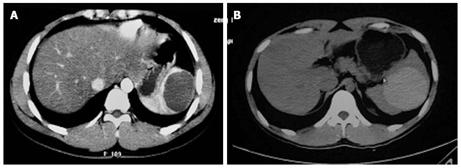
Figure 1 Abdominal computed tomography.
A: Abdominal computed tomography of one case revealed a splenic cyst; B: Abdominal computed tomography of another case revealed a splenic hematoma.
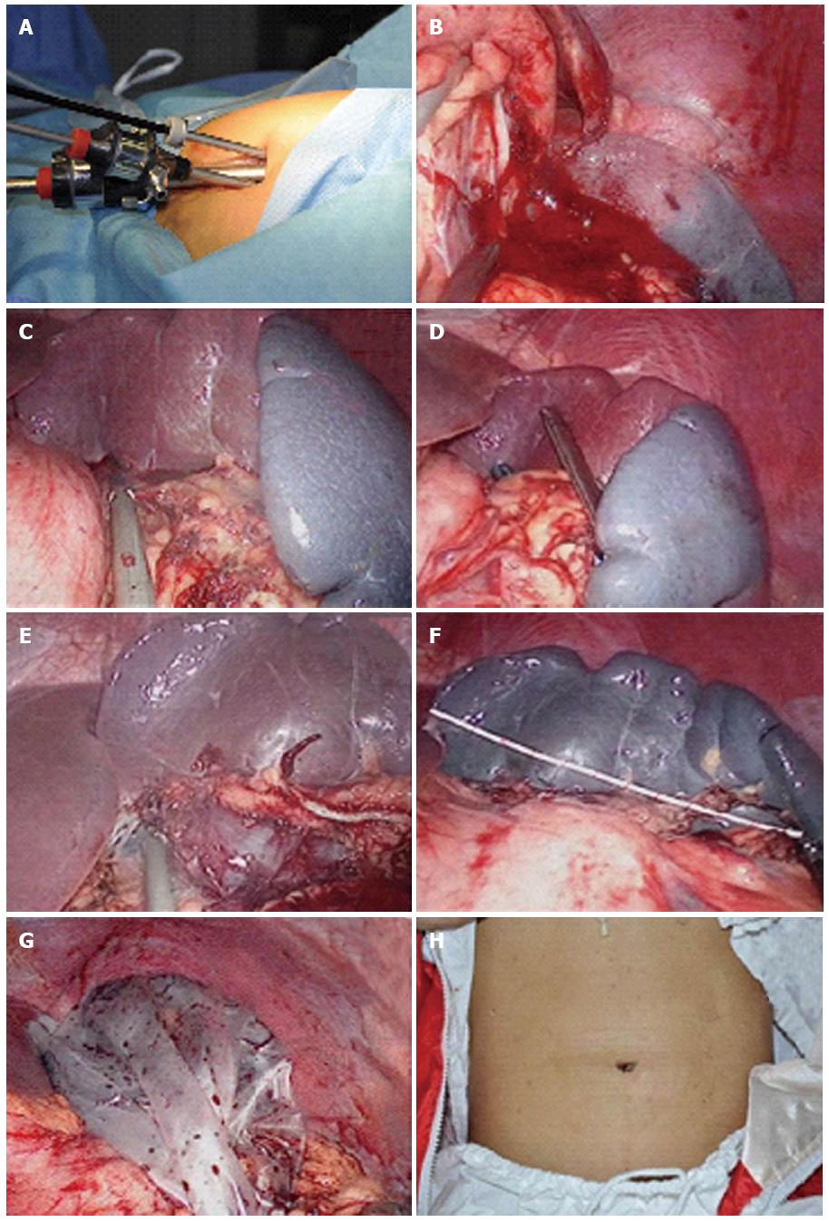
Figure 2 Surgical procedure.
A: The entry ports at the single umbilical incision; B: Resection of the gastrolienal ligament (case 1); C: Resection of the gastrolienal ligament (case 2); D: Cutting of the splenic pedicle with the linear cutting stapler; E: Resection of the phrenicosplenic ligament; F: Measurement of the spleen length; G: Specimen placed into retrieval bag; H: Appearance of wound 1 mo postoperatively.
- Citation: Liang ZW, Cheng Y, Jiang ZS, Liu HY, Gao Y, Pan MX. Transumbilical single-incision endoscopic splenectomy: Report of ten cases. World J Gastroenterol 2014; 20(1): 258-263
- URL: https://www.wjgnet.com/1007-9327/full/v20/i1/258.htm
- DOI: https://dx.doi.org/10.3748/wjg.v20.i1.258


