Copyright
©2013 Baishideng Publishing Group Co.
World J Gastroenterol. Feb 14, 2013; 19(6): 951-954
Published online Feb 14, 2013. doi: 10.3748/wjg.v19.i6.951
Published online Feb 14, 2013. doi: 10.3748/wjg.v19.i6.951
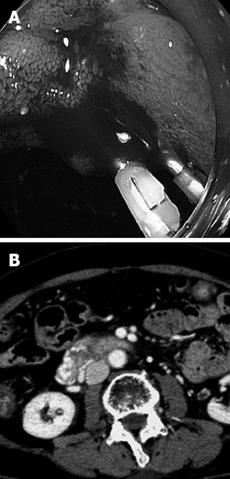
Figure 1 Endoscopy and computed tomography of the duodenum.
A: Endoscopy demonstrates bleeding varices in the second portion of the duodenum; B: Contrast-enhanced computed tomography reveals markedly tortuous varices around the wall in the second and third portion of the duodenum.
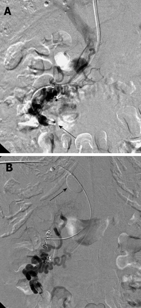
Figure 2 Balloon-occluded retrograde venography.
A: Balloon-occluded retrograde venography (BRTV) shows the dilated efferent vein and the duodenal varices, but the contrast material quickly disappears through several afferent veins. Note that the balloon was inflated in the right ovarian vein (arrow); B: BRTV with occlusion of the main portal trunk (arrow) after embolization of one of the afferent veins reveals the complete opacification of the duodenal varices.
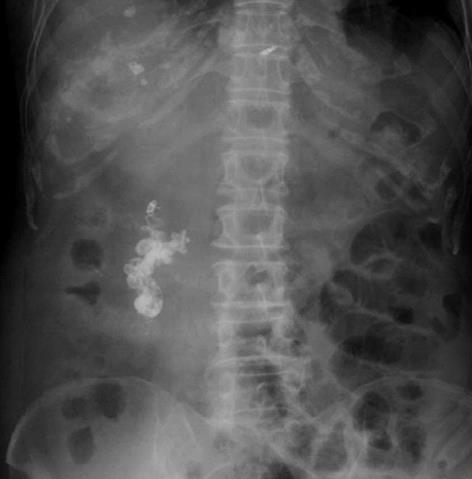
Figure 3 Radiograph after embolization of the duodenal varices demonstrates complete and adequate accumulation of the ethiodized oil in the varices.
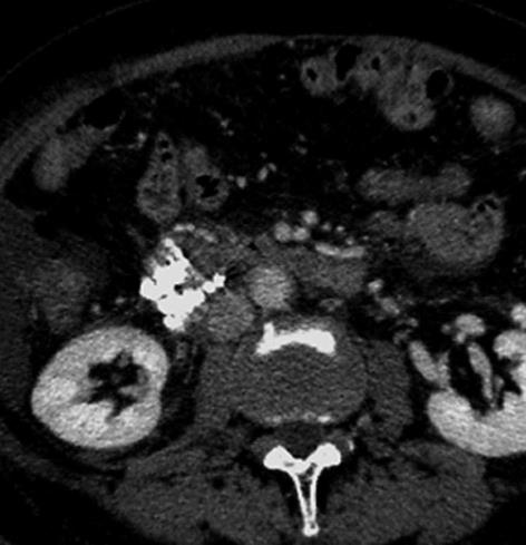
Figure 4 Contrast-enhanced computed tomography after embolization of the duodenal varices shows the complete accumulation of ethiodized oil in the varices.
- Citation: Hashimoto R, Sofue K, Takeuchi Y, Shibamoto K, Arai Y. Successful balloon-occluded retrograde transvenous obliteration for bleeding duodenal varices using cyanoacrylate. World J Gastroenterol 2013; 19(6): 951-954
- URL: https://www.wjgnet.com/1007-9327/full/v19/i6/951.htm
- DOI: https://dx.doi.org/10.3748/wjg.v19.i6.951


