Copyright
©2013 Baishideng Publishing Group Co.
World J Gastroenterol. Dec 28, 2013; 19(48): 9240-9255
Published online Dec 28, 2013. doi: 10.3748/wjg.v19.i48.9240
Published online Dec 28, 2013. doi: 10.3748/wjg.v19.i48.9240
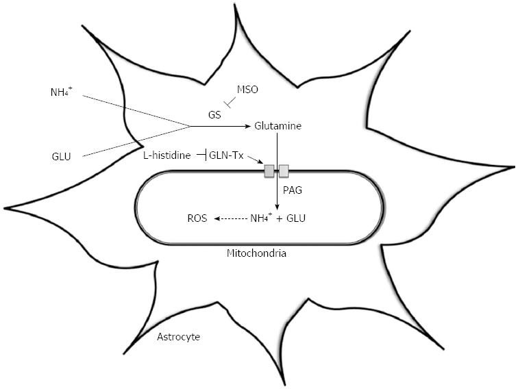
Figure 1 The ‘Trojan Horse’ hypothesis.
This illustrates the synthesis of glutamine via the enzyme glutamine synthetase; its transport into mitochondria via the glutamine transporter (GLN-Tx); its hydrolysis by phosphate-activated glutaminase (PAG) resulting in glutamate (GLU) and ammonia (NH4+) production and the subsequent generation of reactive oxygen species (ROS). MSO: L-methionine S-sulfoximinel; GS: Glutamine synthetase.
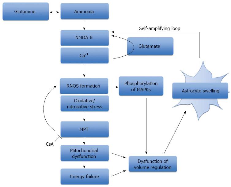
Figure 2 The role of oxidative stress, mitochondrial permeability transition and energy failure in ammonia-induced neurotoxicity.
A schematic representation of the central role that ammonia plays in the production of oxidative/nitrosative stress and astrocyte swelling. Ammonia-induced astrocyte swelling is mediated by oxidative and nitrosative stress resulting in the induction of the MPT, activation of intracellular signaling kinases and alterations in gene expression. Mitochondrial dysfunction and energy failure culminates in astrocytes failing to regulate their cell volume, thereby resulting in astrocyte swelling. NMDA-R: N-methyl-D-aspartate-receptor; RNOS: Reactive nitrogen and oxygen species; MPT: Mitochondrial permeability transition; MAPKs: Mitogen-activated protein kinases; CsA: Cyclosporine A.
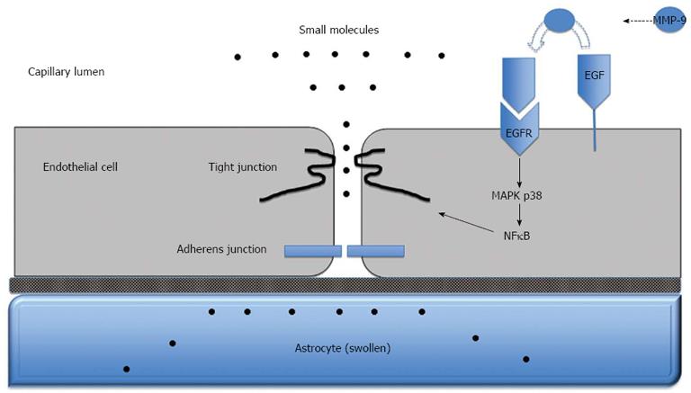
Figure 3 Blood-brain barrier dysfunction in acute liver failure.
Anatomy of the blood-brain barrier (BBB) created by the brain capillary endothelial cell and its paracellular tight junction and adherens junction. In acute liver failure, activation of epidermal growth factor receptor (EGFR) and other signaling pathways results in a loss of BBB tight junction integrity. Tight junctional proteins are altered, resulting in increased permeability to small molecules, leading to astrocyte swelling. MMP-9: Matrix metalloproteinase-9; MAPK p38: Mitogen activated protein kinase p38; NFκB: Nuclear factor-κB.
- Citation: Scott TR, Kronsten VT, Hughes RD, Shawcross DL. Pathophysiology of cerebral oedema in acute liver failure. World J Gastroenterol 2013; 19(48): 9240-9255
- URL: https://www.wjgnet.com/1007-9327/full/v19/i48/9240.htm
- DOI: https://dx.doi.org/10.3748/wjg.v19.i48.9240


