Copyright
©2013 Baishideng Publishing Group Co.
World J Gastroenterol. Dec 7, 2013; 19(45): 8258-8268
Published online Dec 7, 2013. doi: 10.3748/wjg.v19.i45.8258
Published online Dec 7, 2013. doi: 10.3748/wjg.v19.i45.8258
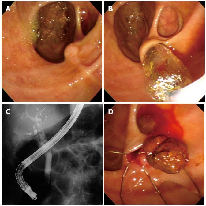
Figure 1 Endoscopic papillary balloon dilation with a small dilating balloon.
A: Huge periampullary diverticulos were noted near the ampulla; B: The 8 mm sized small balloon is gradually inflated with diluted contrast material; inflation is maintained for 30 s; C: Fluoroscopy during balloon dilation shows complete disappearance of the sphincter waist; D: A common bile duct stone was removed by basket through the enlarged biliary orifice.
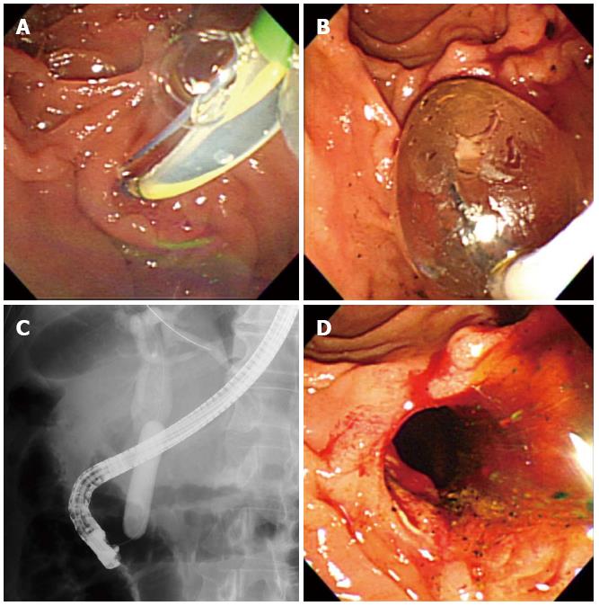
Figure 2 Endoscopic papillary large balloon dilation with minor sphincterotomy.
A: A minor incision of up to one-third of the papilla was performed over a guidewire; B: The 15 mm sized large balloon is gradually inflated with diluted contrast material; inflation is maintained for 30 s; C: Fluoroscopy during balloon dilation shows complete disappearance of the sphincter waist; D: A large biliary orifice can be seen after balloon dilation.
- Citation: Jeong SU, Moon SH, Kim MH. Endoscopic papillary balloon dilation: Revival of the old technique. World J Gastroenterol 2013; 19(45): 8258-8268
- URL: https://www.wjgnet.com/1007-9327/full/v19/i45/8258.htm
- DOI: https://dx.doi.org/10.3748/wjg.v19.i45.8258


