Copyright
©2013 Baishideng Publishing Group Co.
World J Gastroenterol. Dec 7, 2013; 19(45): 8227-8237
Published online Dec 7, 2013. doi: 10.3748/wjg.v19.i45.8227
Published online Dec 7, 2013. doi: 10.3748/wjg.v19.i45.8227
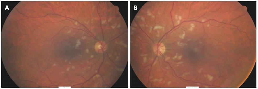
Figure 1 Fundus photographs of a 60-year-old male treated with high dose interferon-α for renal cell carcinoma.
These images show bilateral, typical interferon-associated retinopathy consisting of cotton wool spots and retinal hemorrhages surround the optic disc.
- Citation: O’Day R, Gillies MC, Ahlenstiel G. Ophthalmologic complications of antiviral therapy in hepatitis C treatment. World J Gastroenterol 2013; 19(45): 8227-8237
- URL: https://www.wjgnet.com/1007-9327/full/v19/i45/8227.htm
- DOI: https://dx.doi.org/10.3748/wjg.v19.i45.8227


