Copyright
©2013 Baishideng Publishing Group Co.
World J Gastroenterol. Nov 28, 2013; 19(44): 8047-8055
Published online Nov 28, 2013. doi: 10.3748/wjg.v19.i44.8047
Published online Nov 28, 2013. doi: 10.3748/wjg.v19.i44.8047
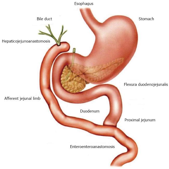
Figure 1 Schematic image of Roux-en-Y hepaticojejunoanastomosis[22].
Figure 2 Endoscopic image.
A: Successful cannulation of a stenotic orifice of the hepaticojejunoanastomosis (HJA) (followed by successful cannulation of adjoining bile ducts); B: Dilation balloon in a deflated form successfully inserted down the guide wire into the area of the stenotic HJA; C: Last phase of a successful endoscopic insertion of a 7 Fr plastic biliary drain into the stenotic HJA and adjoining bile ducts; D: Highly satisfactory final effect of endoscopic treatment of the stenotic HJA (with inserted guide wire).
Figure 3 Endoscopic retrograde cholangiography performed by using single balloon enteroscopy.
A: A patient with hepaticojejunoanastomosis (HJA) shows comparatively narrow HJA stenosis with suprastenotic dilation of bile ducts; B: The patient from Figure 3A in whom HJA stenosis was successfully bridged by endoscopic insertion of a 7 Fr plastic biliary stent.
- Citation: Kianička B, Lata J, Novotný I, Dítě P, Vaníček J. Single balloon enteroscopy for endoscopic retrograde cholangiography in patients with Roux-en-Y hepaticojejunoanastomosis. World J Gastroenterol 2013; 19(44): 8047-8055
- URL: https://www.wjgnet.com/1007-9327/full/v19/i44/8047.htm
- DOI: https://dx.doi.org/10.3748/wjg.v19.i44.8047


