Copyright
©2013 Baishideng Publishing Group Co.
World J Gastroenterol. Nov 21, 2013; 19(43): 7772-7777
Published online Nov 21, 2013. doi: 10.3748/wjg.v19.i43.7772
Published online Nov 21, 2013. doi: 10.3748/wjg.v19.i43.7772
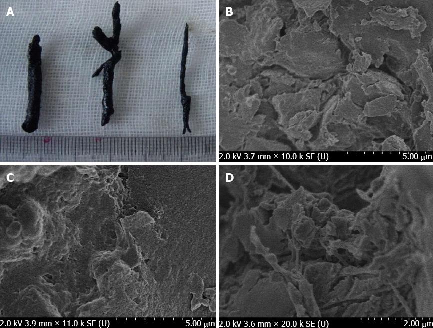
Figure 1 Morphology of biliary casts.
A: Cordlike, columnar and dendritic shapes of biliary casts within the biliary ductal system; B: A biliary cast in the shape of an irregular sheet composed of imbricated accumulation (× 10000); C: A biliary cast in the shape of a honeycombs with porous structures and adherent crystalline substances (× 10000); D: A biliary cast in the shape of filamentous structures (× 10000).
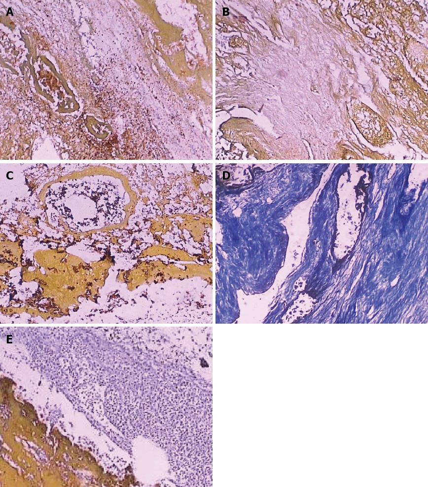
Figure 2 Histological and immunohistochemical examination of biliary casts.
A: Histological examination of a biliary cast, showing tubiform structures (HE staining × 100); B: Histological examination of a biliary cast, showing filamentous structures (HE staining × 100); C: A biliary cast with tubiform structures positive for CD34 (brown color, × 100); D: A biliary cast with filamentous structures positive for collagen fibers (Masson staining × 100); E: A biliary cast showing peripheral positivity for neutrophils and other inflammatory cells (HE staining, × 100).
- Citation: Yang YL, Zhang C, Lin MJ, Shi LJ, Zhang HW, Li JY, Yu Q. Biliary casts after liver transplantation: Morphology and biochemical analysis. World J Gastroenterol 2013; 19(43): 7772-7777
- URL: https://www.wjgnet.com/1007-9327/full/v19/i43/7772.htm
- DOI: https://dx.doi.org/10.3748/wjg.v19.i43.7772


