Copyright
©2013 Baishideng Publishing Group Co.
World J Gastroenterol. Sep 28, 2013; 19(36): 6084-6092
Published online Sep 28, 2013. doi: 10.3748/wjg.v19.i36.6084
Published online Sep 28, 2013. doi: 10.3748/wjg.v19.i36.6084
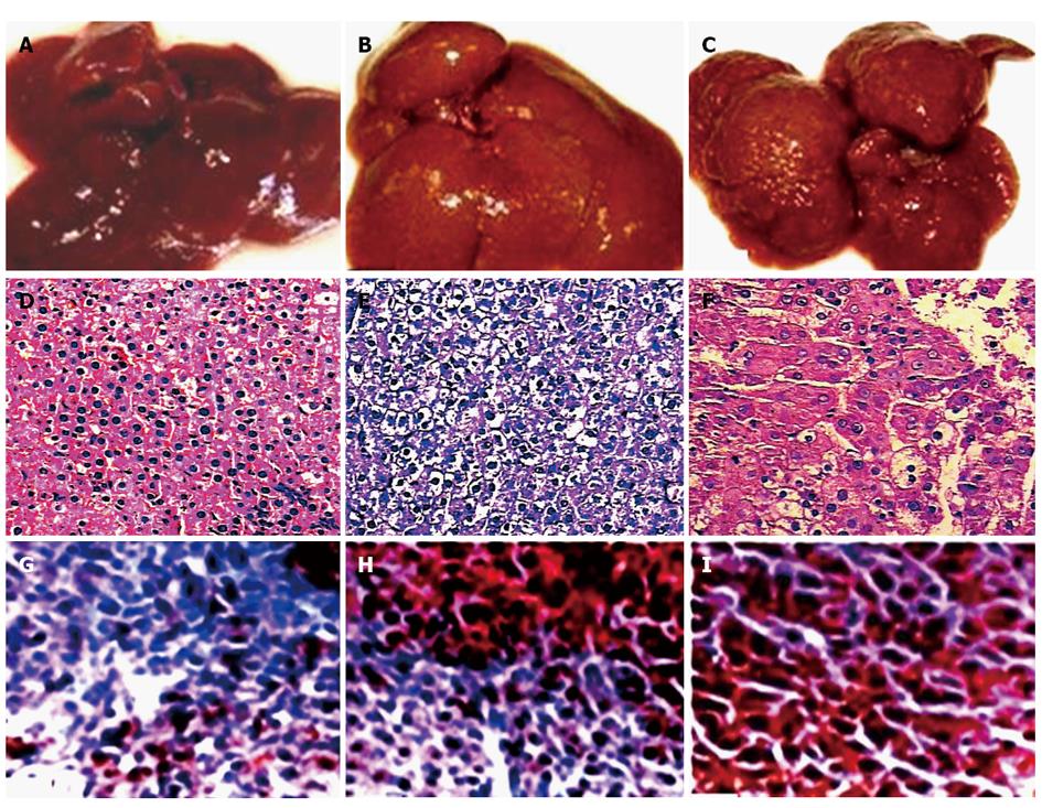
Figure 1 Rat livers and the histopathological alterations of rat tissues during malignant transformation of hepatocytes.
A-C: The appearances of rat livers at different stages at early stage (A), at intermediate stage (B), and at later stage (C); D-F: The histopathology alteration (hematoxylin and eosin staining) of the corresponding rat liver section (× 200); G-I: The immunohistochemical confirmation of corresponding rat hepatic gamma-glutamyl transferase at the same stage (× 200).
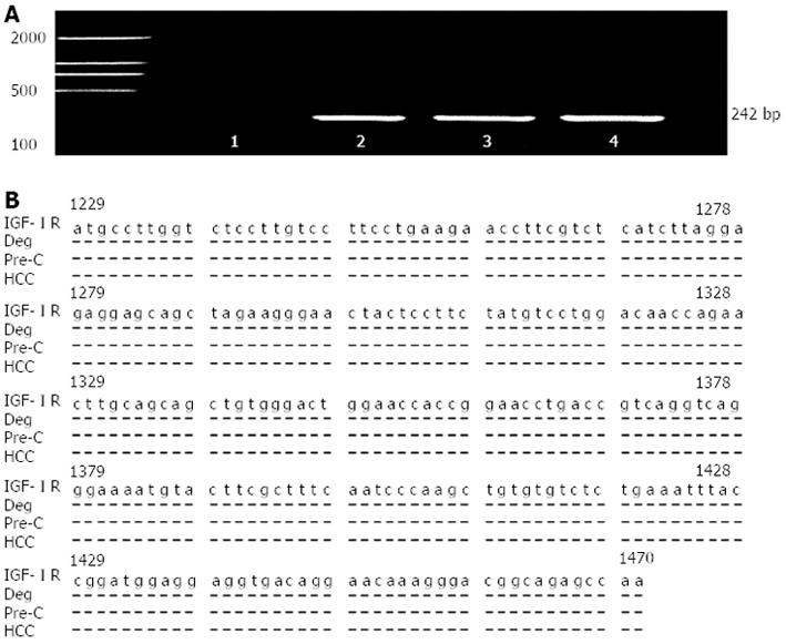
Figure 2 Gene amplification, sequencing and homological analysis of liver insulin-like growth factor I receptor.
A: The amplified fragments of rat insulin-like growth factor I receptor (IGF-IR) mRNA by reverse transcription-nested polymerase chain reaction 1, no positive fragments from control rat liver 2, the positive fragments from degeneration rat (Deg) liver; 3, the positive fragments from precancerous rat (Pre-C) liver, and 4, the positive fragments from hepatocellular carcinoma (HCC) rat liver; B: The amplified fragment of rat IGF-IR mRNA was confirmed by sequencing and alignment of nucleotide sequences from rat liver.
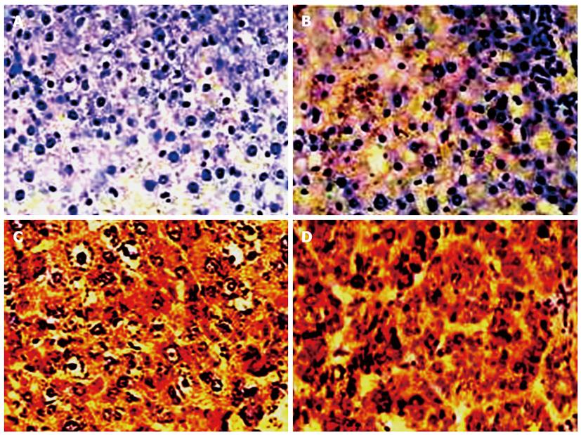
Figure 3 Immunohistochemical analysis of rat liver insulin-like growth factor I receptor at the different stage of rat hepatocyte malignant transformation (× 200).
A: No positive staining in the liver from control rat; B: The weaker insulin-like growth factor I receptor (IGF-IR) expression in the liver from degeneration rat; C: The significantly IGF-IR expression in the liver from precancerous rat; D: The over-expression of liver IGF-IR in hepatoma rats.
- Citation: Yan XD, Yao M, Wang L, Zhang HJ, Yan MJ, Gu X, Shi Y, Chen J, Dong ZZ, Yao DF. Overexpression of insulin-like growth factor-I receptor as a pertinent biomarker for hepatocytes malignant transformation. World J Gastroenterol 2013; 19(36): 6084-6092
- URL: https://www.wjgnet.com/1007-9327/full/v19/i36/6084.htm
- DOI: https://dx.doi.org/10.3748/wjg.v19.i36.6084


