Copyright
©2013 Baishideng Publishing Group Co.
World J Gastroenterol. Sep 21, 2013; 19(35): 5936-5939
Published online Sep 21, 2013. doi: 10.3748/wjg.v19.i35.5936
Published online Sep 21, 2013. doi: 10.3748/wjg.v19.i35.5936
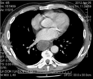
Figure 1 Computed tomography (mediastinal window setting) shows bulky neoformation jutting into the esophageal lumen.
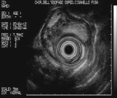
Figure 2 Endoscopic ultrasound shows a solid, hypoechoic, neoformation, with hyperechoic areas and a vascular network.
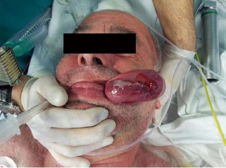
Figure 3 Giant, regurgitated hypopharyngeal polyp.
- Citation: Pallabazzer G, Santi S, Biagio S, D’Imporzano S. Difficult polypectomy-giant hypopharyngeal polyp: Case report and literature review. World J Gastroenterol 2013; 19(35): 5936-5939
- URL: https://www.wjgnet.com/1007-9327/full/v19/i35/5936.htm
- DOI: https://dx.doi.org/10.3748/wjg.v19.i35.5936


