Copyright
©2013 Baishideng Publishing Group Co.
World J Gastroenterol. Sep 14, 2013; 19(34): 5763-5768
Published online Sep 14, 2013. doi: 10.3748/wjg.v19.i34.5763
Published online Sep 14, 2013. doi: 10.3748/wjg.v19.i34.5763
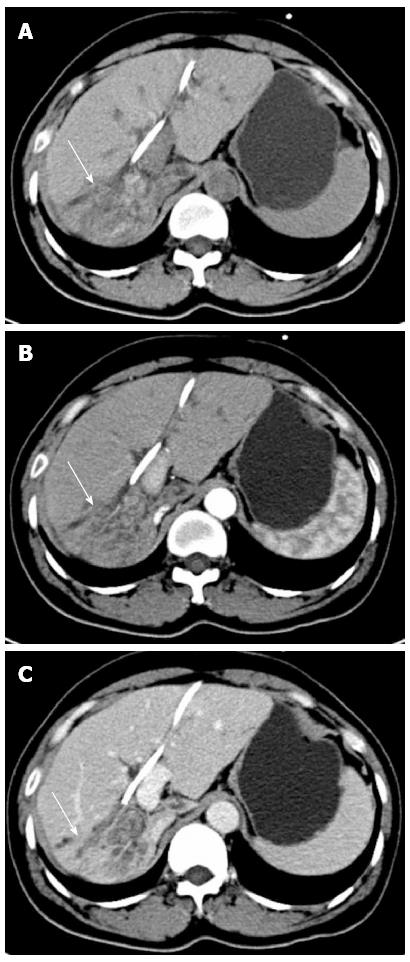
Figure 1 Preoperative computed tomography scan shows hepatolithiasis in the right liver with hepatic parenchymal atrophy (arrow).
A: Non-contrast; B: Arterial phase; C: Venous phase.
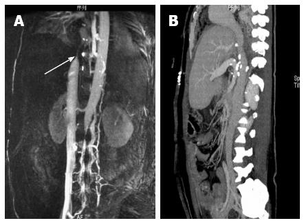
Figure 2 The inferior vena cava.
A: Pretreatment of stenosis in inferior vena cava (IVC) on magnetic resonance venogram imaging (arrow); B: One year after patch repair, computed tomography angiography showed no obvious obstruction in the IVC and good hepatic vein outflow.
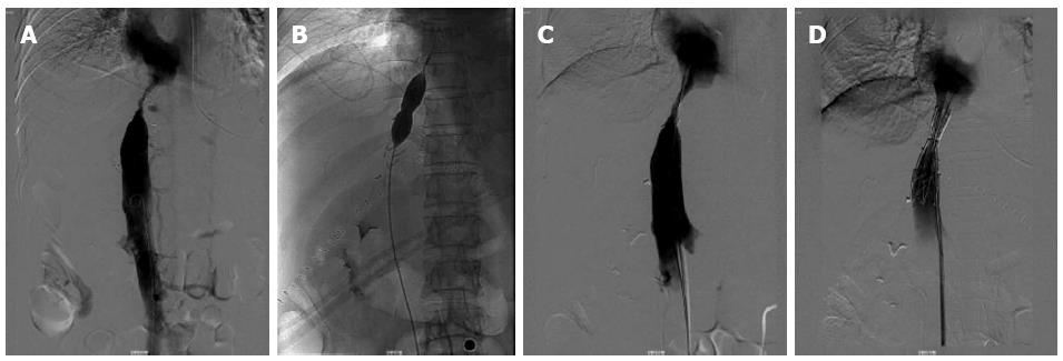
Figure 3 Treating stenosis of the inferior vena cava by balloon dilation and stent placement.
A: Digital subtraction angiography shows the stenosis of inferior vena cava; B: Balloon dilation; C: After balloon angioplasty; D: Metallic stenting.
Figure 4 Diagram of the operative procedure for removing the metallic stent from the inferior vena cava and repairing the stenosis with a BalMedic pericardial patch.
A: The proximal end of the stent was directly at the entrance of the hepatic veins into the inferior vena cava (IVC); B: Open the stenosis of IVC; C: BalMedic pericardial patch was anastomosed to the IVC to broaden its lumen; D: After patch repair of the IVC. LHV: Left hepatic vein; MHV: Middle hepatic vein; RHV: Right hepatic vein.
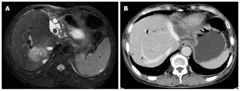
Figure 5 Preoperative imaging findings.
A: Magnetic resonance imaging shows bilateral hepatolithiasis with infection at the posterior right lobe of the liver; B: Major hepatic vein on computed tomography.
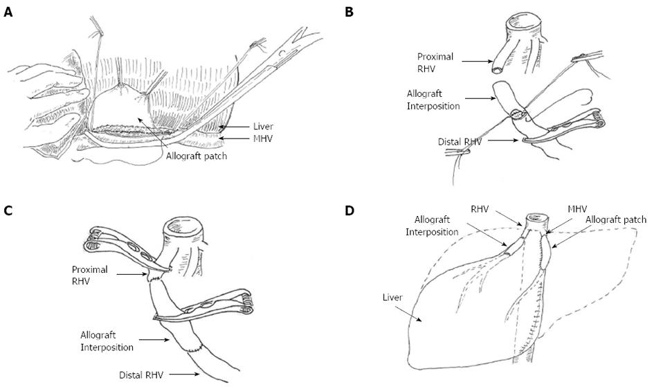
Figure 6 Diagram of reconstruction of middle and right hepatic vein.
A: Middle hepatic vein (MHV) reconstruction using a common iliac artery allogenic graft; B: Right hepatic vein (RHV) reconstruction using an allogeneic iliac vein graft; C: Completion of RHV reconstruction using allograft interposition; D: Accomplishment of MHV and RHV outflow.
Figure 7 Postoperative computed tomography scan.
A, B: Demonstrated good right hepatic vein tract; C: The inter-positioned graft using an allogeneic iliac vein (arrow).
- Citation: Bai XL, Chen YW, Zhang Q, Ye LY, Xu YL, Wang L, Cao CH, Gao SL, Khoodoruth MAS, Ramjaun BZ, Dong AQ, Liang TB. Acute iatrogenic Budd-Chiari syndrome following hepatectomy for hepatolithiasis: A report of two cases. World J Gastroenterol 2013; 19(34): 5763-5768
- URL: https://www.wjgnet.com/1007-9327/full/v19/i34/5763.htm
- DOI: https://dx.doi.org/10.3748/wjg.v19.i34.5763


