Copyright
©2013 Baishideng Publishing Group Co.
World J Gastroenterol. Sep 14, 2013; 19(34): 5593-5597
Published online Sep 14, 2013. doi: 10.3748/wjg.v19.i34.5593
Published online Sep 14, 2013. doi: 10.3748/wjg.v19.i34.5593
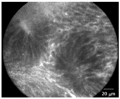
Figure 1 Rectal mucosa of patient in remission from ulcerative colitis.
Irregular alignment of crypt, crypt distortion and fusion with reduced amount of goblet cells.
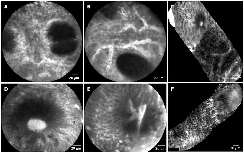
Figure 2 Colonic mucosa.
A: Patient in remission from ulcerative colitis. Crypt distortion and fusion and many capillaries visible in the lamina propria; B: Patient in remission from ulcerative colitis. Enlarged spaces between crypts and dilated prominent branching vessels; C: In patient with active ulcerative colitis (distal colitis). Image of colonic mucosa showing the switch from normal mucosa (top of the figure) to inflamed mucosa Inflamed mucosa showing irregular arrangement of crypts, crypt fusion and capillaries alterations; D: In patient with active ulcerative colitis. Dilated and bright crypt lumen (fluorescein leakage) with intact epithelium; E: In patient with active ulcerative colitis. Dilated, irregular and bright crypt lumen (fluorescein leakage) with partially intact epithelium; F: In patient with highly active ulcerative colitis (Mayo CU3). Crypts distortion and destruction, crypt abscess and crypts replacement by diffuse necrosis.
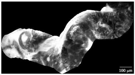
Figure 3 Dysplasia-associated lesional mass in long-standing ulcerative colitis.
Image of colonic mucosa evidencing the switch from the inflamed mucosa, to the neoplastic mucosa. Inflamed mucosa is characterized by crypts fusion and distortion, dilation of crypt openings, enlarged spaces between crypts, and microvascular alterations with fluorescein leaks into the crypt lumen. Dysplastic mucosa (right corner) is characterized by “dark” cells, irregular architectural patterns with villiform structures and a “dark” epithelial border.
- Citation: Palma GDD, Rispo A. Confocal laser endomicroscopy in inflammatory bowel diseases: Dream or reality? World J Gastroenterol 2013; 19(34): 5593-5597
- URL: https://www.wjgnet.com/1007-9327/full/v19/i34/5593.htm
- DOI: https://dx.doi.org/10.3748/wjg.v19.i34.5593


