Copyright
©2013 Baishideng Publishing Group Co.
World J Gastroenterol. Sep 7, 2013; 19(33): 5557-5564
Published online Sep 7, 2013. doi: 10.3748/wjg.v19.i33.5557
Published online Sep 7, 2013. doi: 10.3748/wjg.v19.i33.5557
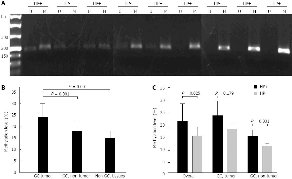
Figure 1 Detection of transforming growth factor-β1 promoter methylation by methylation-specific polymerase chain reaction.
Genomic DNA was extracted from human gastric tissues. A: Representative methylation-specific polymerase chain reaction results from gastric cancer (GC) tissues with or without Helicobacter pylori (H. pylori) infection are shown; B: Levels of transforming growth factor-β1 (TGF-β1) promoter methylation in GC tissues, non-cancerous gastric mucosa from the GC patients (GC, non-tumor) and normal gastric mucosa from non-GC subjects (Non-GC tissues); C: Impact of H. pylori status on the levels of TGF-β1 promoter methylation in GC tissues (GC, tumor), non-cancerous gastric mucosa from the GC patients (GC, non-tumor) and combined samples (overall). HP+: H. pylori-positive; HP-: H. pylori-negative; U: Unmethylated; M: Methylated.
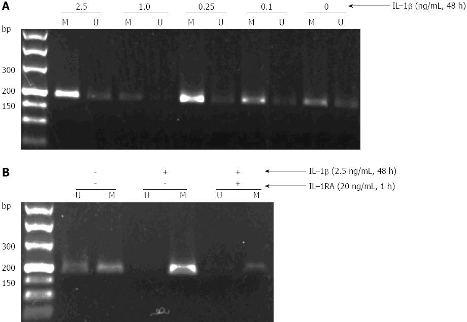
Figure 2 Induction of transforming growth factor-β1 methylation by interleukin-1β in GES-1 cells.
A: Treatment of GES-1 cells by interleukin (IL)-1β led to a dose-dependent methylation of transforming growth factor (TGF)-β1; B: IL-1β-induced TGF-β1 methylation in GES-1cells was partially abolished by IL-1RA. U: Unmethylated; M: Methylated; H. pylori: Helicobacter pylori.
-
Citation: Wang YQ, Li YM, Li X, Liu T, Liu XK, Zhang JQ, Guo JW, Guo LY, Qiao L. Hypermethylation of
TGF-β1 gene promoter in gastric cancer. World J Gastroenterol 2013; 19(33): 5557-5564 - URL: https://www.wjgnet.com/1007-9327/full/v19/i33/5557.htm
- DOI: https://dx.doi.org/10.3748/wjg.v19.i33.5557


