Copyright
©2013 Baishideng Publishing Group Co.
World J Gastroenterol. Aug 28, 2013; 19(32): 5389-5392
Published online Aug 28, 2013. doi: 10.3748/wjg.v19.i32.5389
Published online Aug 28, 2013. doi: 10.3748/wjg.v19.i32.5389
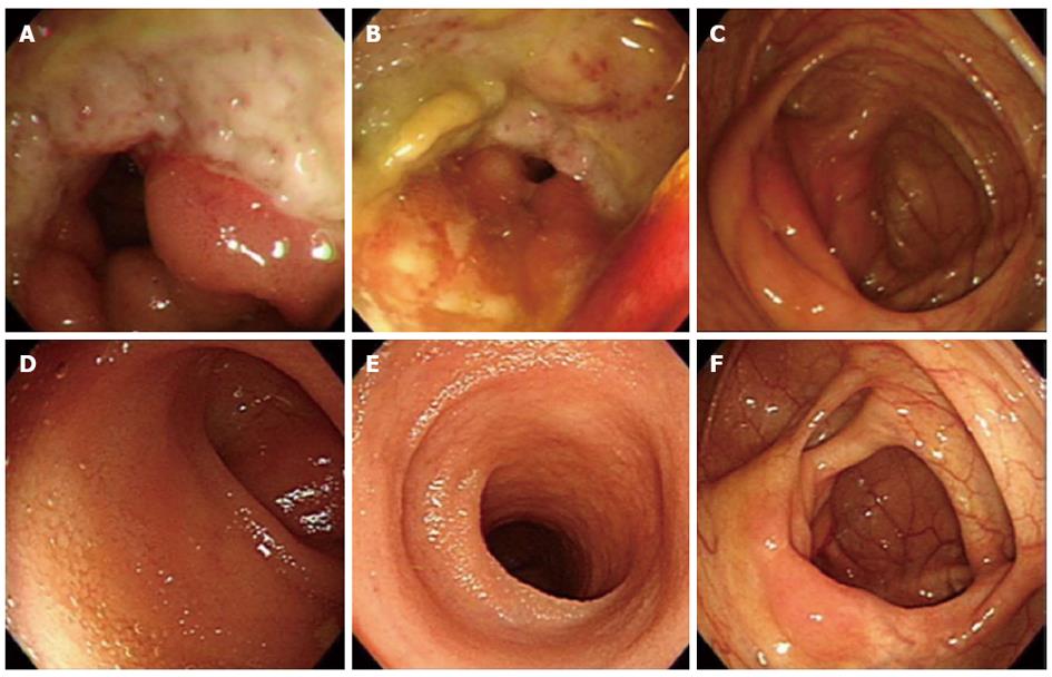
Figure 1 A colonoscopy on admission revealed a large, deep, well-demarcated ulcer with exudate, mucosal edema and erythema at the terminal ileum (A-C).
On follow-up colonoscopy at 36 mo, the ulcer at the terminal ileum was replaced by normal mucosa (D-F) with complete mucosal healing.
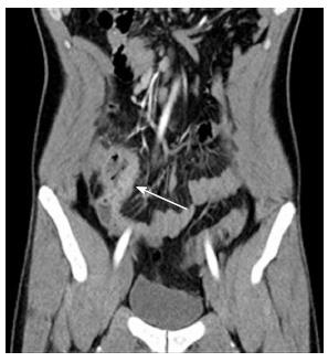
Figure 2 In computed tomography, arrow showed bowel wall thickening and prominent enhancement with surrounding fat infiltration at the terminal ileum and cecum.
This was suggestive of active inflammation.
- Citation: Chung SH, Park SJ, Hong SP, Cheon JH, Kim TI, Kim WH. Intestinal Behçet’s disease appearing during treatment with adalimumab in a patient with ankylosing spondylitis. World J Gastroenterol 2013; 19(32): 5389-5392
- URL: https://www.wjgnet.com/1007-9327/full/v19/i32/5389.htm
- DOI: https://dx.doi.org/10.3748/wjg.v19.i32.5389


