Copyright
©2013 Baishideng Publishing Group Co.
World J Gastroenterol. Aug 14, 2013; 19(30): 5016-5020
Published online Aug 14, 2013. doi: 10.3748/wjg.v19.i30.5016
Published online Aug 14, 2013. doi: 10.3748/wjg.v19.i30.5016
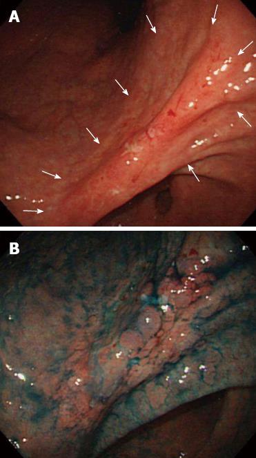
Figure 1 Endoscopic findings upon initial admission for early gastric cancer.
A: Conventional endoscopy showing a superficial depressed type lesion, 50 mm in size with an ulcer scar located in the gastric angle (arrows); B: Chromoendoscopy with indigo-carmine dye (blue) showing the lesion margin.
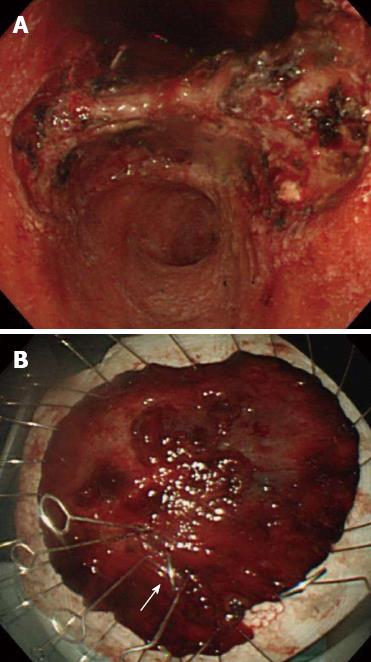
Figure 2 In situ locale of the lesion and gross appearance of the resected tumor.
A: Intraoperative image taken during the endoscopic submucosal dissection (ESD) procedure. ESD was performed in October 2001; B: A tear (arrow) was present inside the lesion and likely was incident to the ESD procedure.
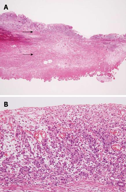
Figure 3 Histological findings of the endoscopic submucosal dissection-resected lesion tissues.
A: The tumor is confined to the mucosa (solid arrow) and an ulcer scar (dashed arrow) (magnification: × 20); B: The tumor is composed of poorly differentiated adenocarcinoma with signet ring cells (magnification: × 100).
Figure 4 No endoscopic evidence of recurrence was found at the endoscopic submucosal dissection site eight years after endoscopic submucosal dissection.
A: Conventional endoscopy; B: Chromoendoscopy with indigo-carmine dye. Endoscopic examination provided no indications of recurrence at the endoscopic submucosal dissection site.
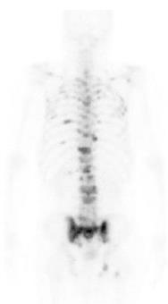
Figure 5 Bone scintigraphy results from the eight-year follow-up showing increased uptake in the spine and pelvic bone.
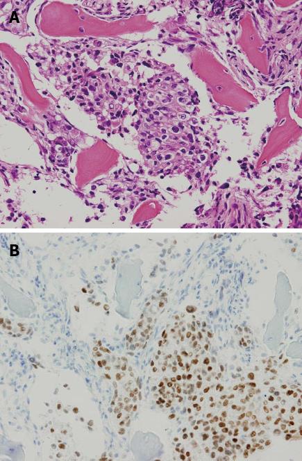
Figure 6 Histological findings of the biopsy specimen taken from the lumbar spine.
A: The biopsy specimen taken from the lumbar spine revealed a poorly differentiated adenocarcinoma histologically resembling the carcinoma cells in the early gastric cancer treated previously by endoscopic submucosal dissection; B: Using immunohistochemical staining, the tumor cells were positive for caudal-type homeobox transcription factor 2.
- Citation: Kawabata H, Oda I, Suzuki H, Nonaka S, Yoshinaga S, Katai H, Taniguchi H, Kushima R, Saito Y. Bone metastasis from early gastric cancer following non-curative endoscopic submucosal dissection. World J Gastroenterol 2013; 19(30): 5016-5020
- URL: https://www.wjgnet.com/1007-9327/full/v19/i30/5016.htm
- DOI: https://dx.doi.org/10.3748/wjg.v19.i30.5016


