Copyright
©2013 Baishideng Publishing Group Co.
World J Gastroenterol. Jul 28, 2013; 19(28): 4624-4629
Published online Jul 28, 2013. doi: 10.3748/wjg.v19.i28.4624
Published online Jul 28, 2013. doi: 10.3748/wjg.v19.i28.4624
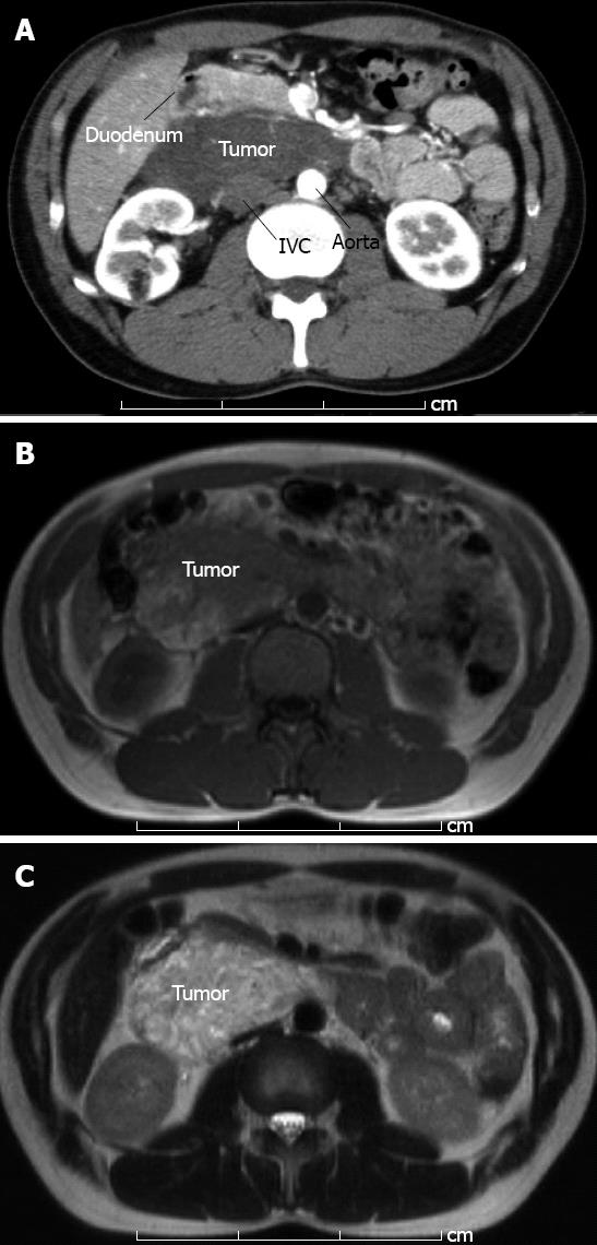
Figure 1 12 cm × 9 cm tumor was detected by computed tomography and magnetic resonance imaging.
A: Abdominal enhanced computed tomography in early phase showed tumor without marked contrast. The tumor had pushed the pancreas to the ventral side; B: T1-weighted image of magnetic resonance imaging showed low and relatively high intensity area inside the tumor; C: T2-weighted image showed high intensity area with a few part of intermediate signal intensity area. IVC: Inferior vena cava.
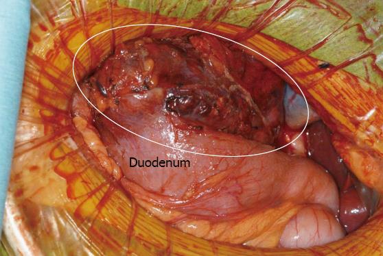
Figure 2 Macroscopic view of intraoperative findings.
The mass was attached to both the duodenum and the pancreas head and required surgical resection by pylorus preserving pancreaticoduodenectomy. The circle denotes the tumor.
Figure 3 Macroscopic findings of the resected specimen.
A: A 120 mm × 95 mm × 50 mm tumor was resected. Scale bar, 70 mm; B: The tumor contained multi-oculated cysts containing intra-cystic hemorrhages.
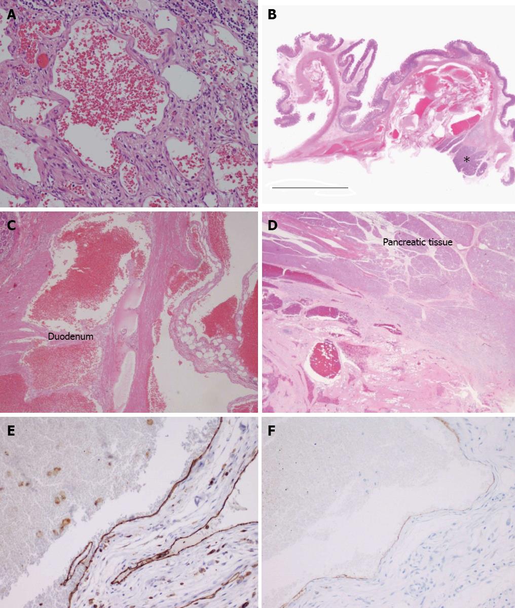
Figure 4 Pathological analysis by hematoxylin and eosin staining and immunohistochemistry.
A: A representative tissue section from the main region of the cavernous hemangioma [hematoxylin and eosin (HE), magnification × 200]; B: The loupe view demonstrates a portion of cavernous hemangioma infiltrating the pancreatic head (asterisk) and duodenal wall (HE). The scale represents 10 mm; C: Tumor invasion into the muscle layer of the duodenum (HE, magnification × 4); D: Tumor invasion into the pancreas head (HE, magnification × 1); E: Positive immunostaining for CD31 supports the diagnosis of hemangioma (magnification × 20); F: The lumen showed partial and weakly positive staining for podoplanin/D2-40 (magnification × 20).
- Citation: Hanaoka M, Hashimoto M, Sasaki K, Matsuda M, Fujii T, Ohashi K, Watanabe G. Retroperitoneal cavernous hemangioma resected by a pylorus preserving pancreaticoduodenectomy. World J Gastroenterol 2013; 19(28): 4624-4629
- URL: https://www.wjgnet.com/1007-9327/full/v19/i28/4624.htm
- DOI: https://dx.doi.org/10.3748/wjg.v19.i28.4624


