Copyright
©2013 Baishideng Publishing Group Co.
World J Gastroenterol. Jul 28, 2013; 19(28): 4531-4536
Published online Jul 28, 2013. doi: 10.3748/wjg.v19.i28.4531
Published online Jul 28, 2013. doi: 10.3748/wjg.v19.i28.4531
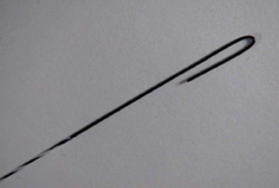
Figure 1 Newly designed J-shaped tip guidewire.
The shape of the tip is a radius of 1 mm; 50 mm from the tip is the start of a hydrophilic coating.
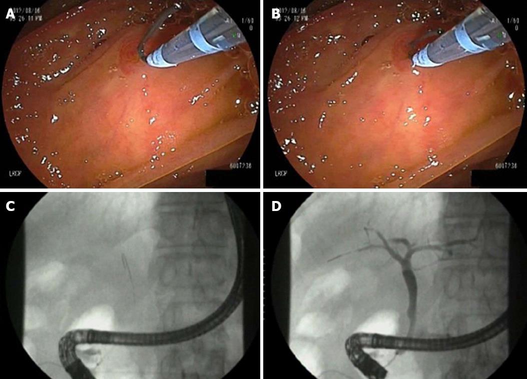
Figure 2 Endoscopic and fluoroscopic images showing the technique with J-shaped tip guidewire.
A: Assistant endoscopist extended approximately 5 mm of the guidewire tip and restored it to the original “J” configuration (stand-by position); B: Selective biliary cannulation was attempted under endoscopic control without fluoroscopy; C: The guidewire was moved in an in-and-out motion by an assisting endoscopist. Once the guidewire was advanced without resistance, fluoroscopy was used to confirm successful cannulation; D: Contrast medium was injected after confirmation of successful biliary cannulation.
Figure 3 An image via intra-duodenal biliary segments of J-shaped tip guidewire (A) and standard guidewire (B).
A: Blunted J-shaped tip may facilitate passage through intra-duodenal segment (arrow); B: Normal guidewire tips may be become stuck in epithelial folds or flexion of intra-duodenal biliary segments (arrowheads).
- Citation: Omuta S, Maetani I, Shigoka H, Gon K, Saito M, Tokuhisa J, Naruki M. Newly designed J-shaped tip guidewire: A preliminary feasibility study in wire-guided cannulation. World J Gastroenterol 2013; 19(28): 4531-4536
- URL: https://www.wjgnet.com/1007-9327/full/v19/i28/4531.htm
- DOI: https://dx.doi.org/10.3748/wjg.v19.i28.4531


