Copyright
©2013 Baishideng Publishing Group Co.
World J Gastroenterol. Jul 21, 2013; 19(27): 4427-4431
Published online Jul 21, 2013. doi: 10.3748/wjg.v19.i27.4427
Published online Jul 21, 2013. doi: 10.3748/wjg.v19.i27.4427
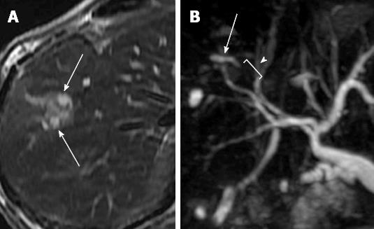
Figure 1 Magnetic resonance imaging.
A: The T2-weighted magnetic resonance imaging shows a hyperintense lesion along the dilated intrahepatic duct (arrows); B: Magnetic resonance cholangiopancreatography shows a stricture of biliary duct (arrow) with the upstream dilatation (arrowhead).
Figure 2 Endoscopic images during cannulation and transpancreatic sphincterotomy.
A: Selective biliary cannulation was attempted in the position of the papilla moved to the lower endoscopic field of view; B: The wire-guided transpancreatic sphincterotomy was performed toward the biliary direction; C: The bile duct orifice was exposed after the transpancreatic sphincterotomy (arrow).
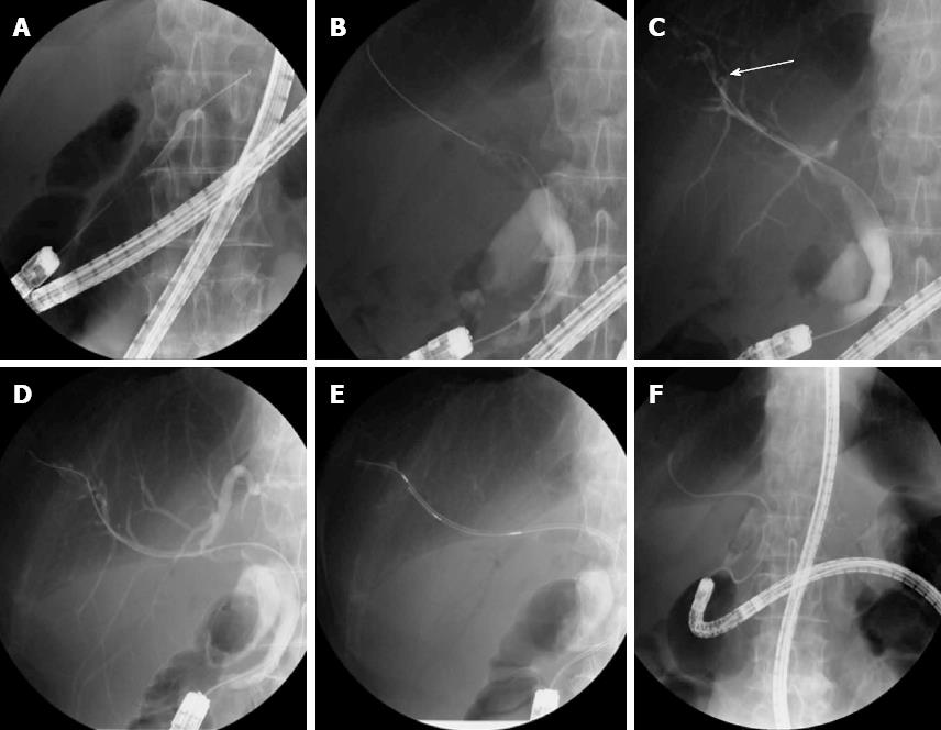
Figure 3 Endoscopic retrograde cholangiopancreatography.
A: A guide wire was inserted deeply into the main pancreatic duct to perform the pre-cutting; B: After the pre-cutting, selective biliary cannulation was achieved; C: Cholangiography revealed a focal stricture in the branch of the right intrahepatic duct (arrow); D: A guide wire was passed through the biliary stricture; E: The brush cytological examination was carried out at the stricture; F: The placement of nasobiliary drainage tube was performed to collect the bile for cytology.
-
Citation: Ikeura T, Shimatani M, Takaoka M, Matsushita M, Miyoshi H, Kurishima A, Sumimoto K, Miyamoto S, Okazaki K. Intrahepatic cholangiocarcinoma diagnosed
via endoscopic retrograde cholangiopancreatography with a short double-balloon enteroscope. World J Gastroenterol 2013; 19(27): 4427-4431 - URL: https://www.wjgnet.com/1007-9327/full/v19/i27/4427.htm
- DOI: https://dx.doi.org/10.3748/wjg.v19.i27.4427


