Copyright
©2013 Baishideng Publishing Group Co.
World J Gastroenterol. Jul 21, 2013; 19(27): 4409-4412
Published online Jul 21, 2013. doi: 10.3748/wjg.v19.i27.4409
Published online Jul 21, 2013. doi: 10.3748/wjg.v19.i27.4409
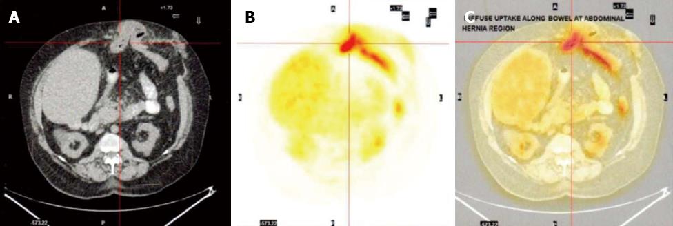
Figure 1 Computed tomography scan (A), flurodeoxyglucose positron emmision tomography (B) and combined computed tomography positron emission tomography image (C) across the site of incisional hernia.
A: Incarcerated hernia with a loop of bowel; B: Confirms “hot spot” with increased flurodeoxyglucose uptake which corresponds with the centre of the incarcerated bowel loop within the hernia with some activity along the efferent loop distal to the hernia.
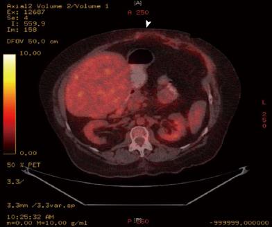
Figure 2 Computed tomography positron emission tomography image 12 mo following the incisional hernia repair.
The arrow heads points to the previous hernia site now repaired anatomically and the “hot spot” has now disappeared.
- Citation: Evans JD, Perera MTP, Pal C, Neuberger J, Mirza DF. Late post liver transplant protein losing enteropathy: Rare complication of incisional hernia. World J Gastroenterol 2013; 19(27): 4409-4412
- URL: https://www.wjgnet.com/1007-9327/full/v19/i27/4409.htm
- DOI: https://dx.doi.org/10.3748/wjg.v19.i27.4409


