Copyright
©2013 Baishideng Publishing Group Co.
World J Gastroenterol. Jul 14, 2013; 19(26): 4185-4191
Published online Jul 14, 2013. doi: 10.3748/wjg.v19.i26.4185
Published online Jul 14, 2013. doi: 10.3748/wjg.v19.i26.4185
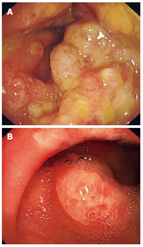
Figure 1 Colonoscopy images of 2 patients with cap polyposis.
A: Colonoscopy image of a patient showing multiple small sessile polypoid lesions with mucous exudates of cap polyposis (CP) in the rectum; B: Colonoscopy image of a patient showing a single sessile red polypoid lesion located on the transverse folds with normal intervening mucosa.
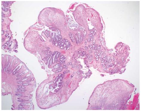
Figure 2 Histology of a sessile colonic polyp from a patient with cap polyposis.
The polyp was comprised of granulation tissue and focally distended crypts with a slightly serrated luminal surface. Surface ulceration with fibrino-mucoid exudates was also present.
- Citation: Li JH, Leong MY, Phua KB, Low Y, Kader A, Logarajah V, Ong LY, Chua JH, Ong C. Cap polyposis: A rare cause of rectal bleeding in children. World J Gastroenterol 2013; 19(26): 4185-4191
- URL: https://www.wjgnet.com/1007-9327/full/v19/i26/4185.htm
- DOI: https://dx.doi.org/10.3748/wjg.v19.i26.4185


