Copyright
©2013 Baishideng Publishing Group Co.
World J Gastroenterol. Jul 7, 2013; 19(25): 4039-4044
Published online Jul 7, 2013. doi: 10.3748/wjg.v19.i25.4039
Published online Jul 7, 2013. doi: 10.3748/wjg.v19.i25.4039
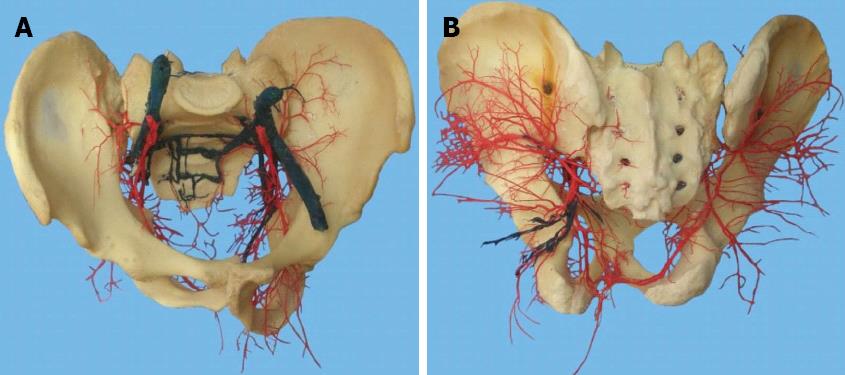
Figure 1 Presacral vascular cast.
A: Front view; B: Dorsal view.
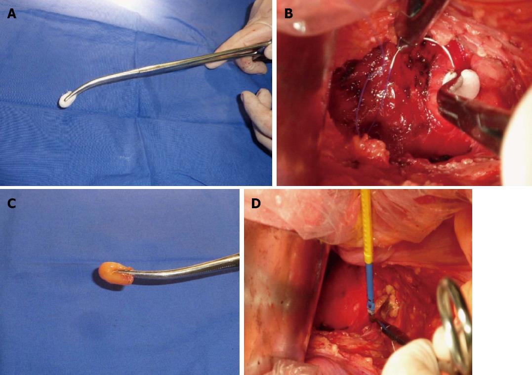
Figure 2 Bleeding point originated from the presacral venous plexus and a sacral neural foramen where the basivertebral veins were injured.
A: Continuous pressure over the bleeding site using a gauze nut at the tip of a long Kelly clamp; B: Venous branches surrounding the gauze nut could be identified, and were suture ligated one by one with 3-0 suture thread; C: Continuous pressure over the bleeding site using the epiploic appendices at the tip of a long Kelly clamp; D: Electrocautery applied through the epiploic appendices pressed with a long Kelly clamp over the bleeding vessel.
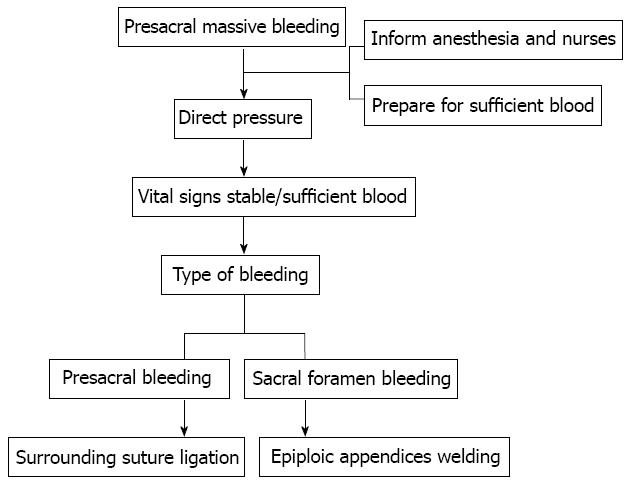
Figure 3 Process in the management of massive presacral bleeding.
- Citation: Lou Z, Zhang W, Meng RG, Fu CG. Massive presacral bleeding during rectal surgery: From anatomy to clinical practice. World J Gastroenterol 2013; 19(25): 4039-4044
- URL: https://www.wjgnet.com/1007-9327/full/v19/i25/4039.htm
- DOI: https://dx.doi.org/10.3748/wjg.v19.i25.4039


