Copyright
©2013 Baishideng Publishing Group Co.
World J Gastroenterol. Jun 28, 2013; 19(24): 3911-3914
Published online Jun 28, 2013. doi: 10.3748/wjg.v19.i24.3911
Published online Jun 28, 2013. doi: 10.3748/wjg.v19.i24.3911
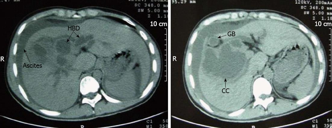
Figure 1 Abdominal computed tomography shows ascites, a normal-sized gallbladder, dilatation of the intrahepatic bile duct, and a huge choledochal cyst.
GB: Gallbladder; HBD: Intrahepatic bile duct; CC: Choledochal cyst.
Figure 2 Abdominal computed tomography shows a rupture in the right wall of the choledochal cyst with hemorrhage (arrow).
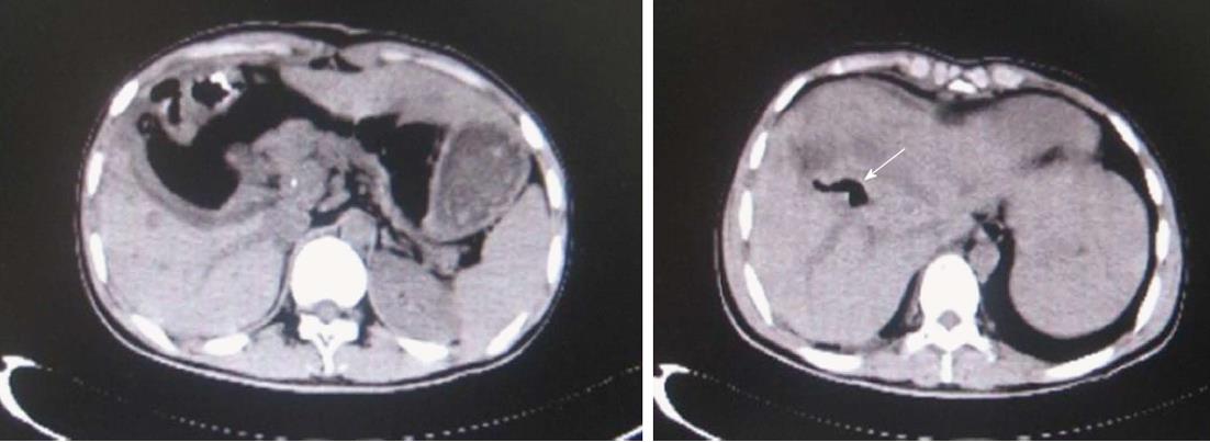
Figure 3 Abdominal computed tomography shows pneumatosis in the intrahepatic bile duct after cyst excision, cholecystectomy and Roux-en-Y hepaticojejunostomy reconstruction (white arrow).
The intrahepatic dilatation tends to reduce in size following sufficient biliary drainage.
- Citation: Duan YF, Yang B, Zhu F. Traumatic rupture of a type IVa choledochal cyst in an adult male. World J Gastroenterol 2013; 19(24): 3911-3914
- URL: https://www.wjgnet.com/1007-9327/full/v19/i24/3911.htm
- DOI: https://dx.doi.org/10.3748/wjg.v19.i24.3911


