Copyright
©2013 Baishideng Publishing Group Co.
World J Gastroenterol. May 21, 2013; 19(19): 2883-2893
Published online May 21, 2013. doi: 10.3748/wjg.v19.i19.2883
Published online May 21, 2013. doi: 10.3748/wjg.v19.i19.2883
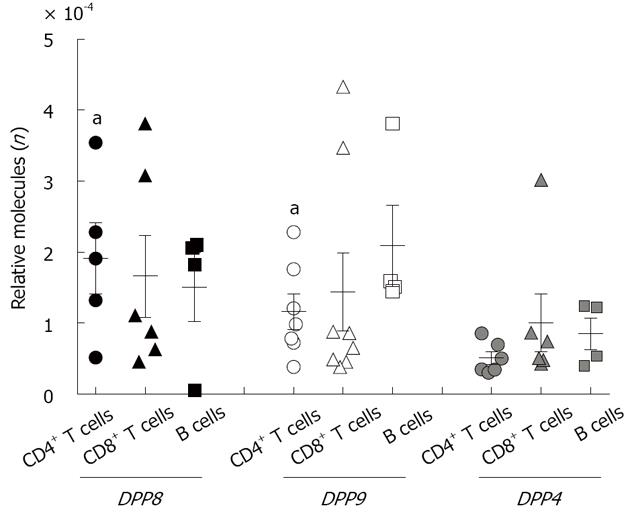
Figure 1 Dipeptidyl peptidase mRNA expression in C57BL/6 mouse splenic lymphocyte subpopulations.
Number of molecules relative to 18S RNA (n = 4-6 mice). aP < 0.05 vs dipeptidyl peptidase (DPP) 4.
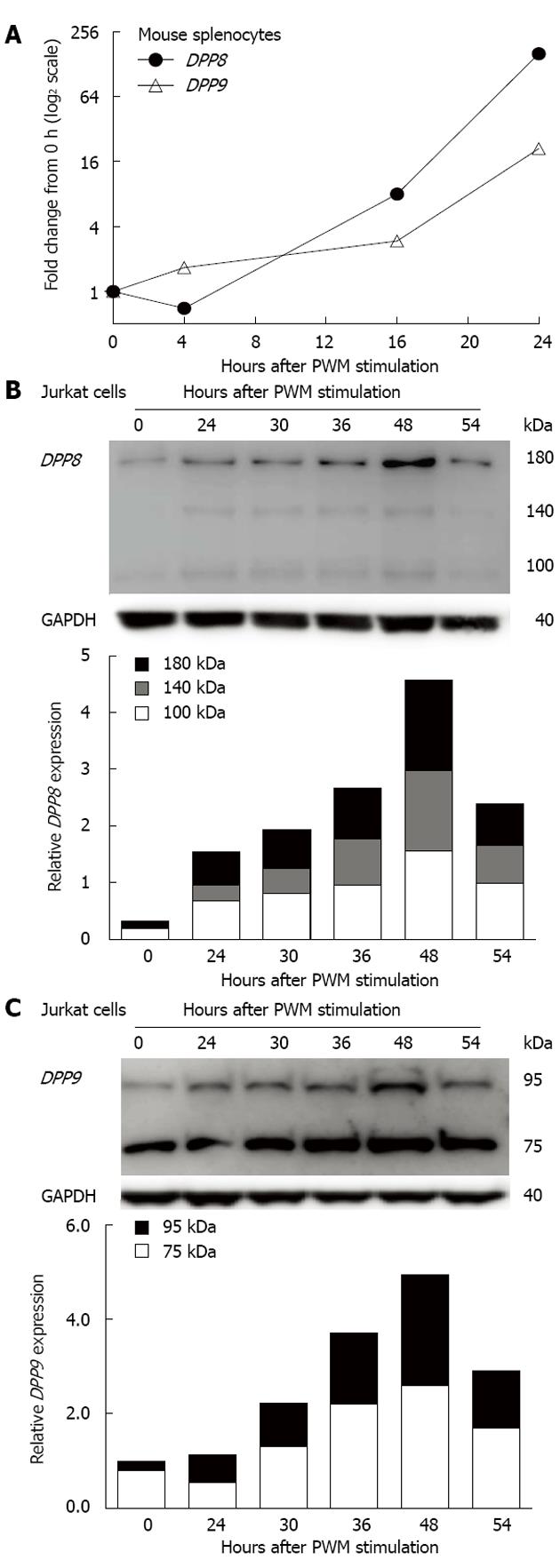
Figure 2 Dipeptidyl peptidase 8 and dipeptidyl peptidase 9 upregulation in pokeweed mitogen stimulated lymphocytes.
A: Dipeptidyl peptidase (DPP) 8 and DPP9 mRNA in mouse splenocytes (representative data from one of three mice); DPP8 and DPP9 proteins from Jurkat cells; B: Immunoblot of DPP8 and densitometry analysis of DPP8 bands; C: Immunoblot of DPP9 and densitometry analysis of bands. Densitometry data shown are relative to glyceraldehyde 3-phosphate dehydrogenase (GAPDH). PWM: Pokeweed mitogen.
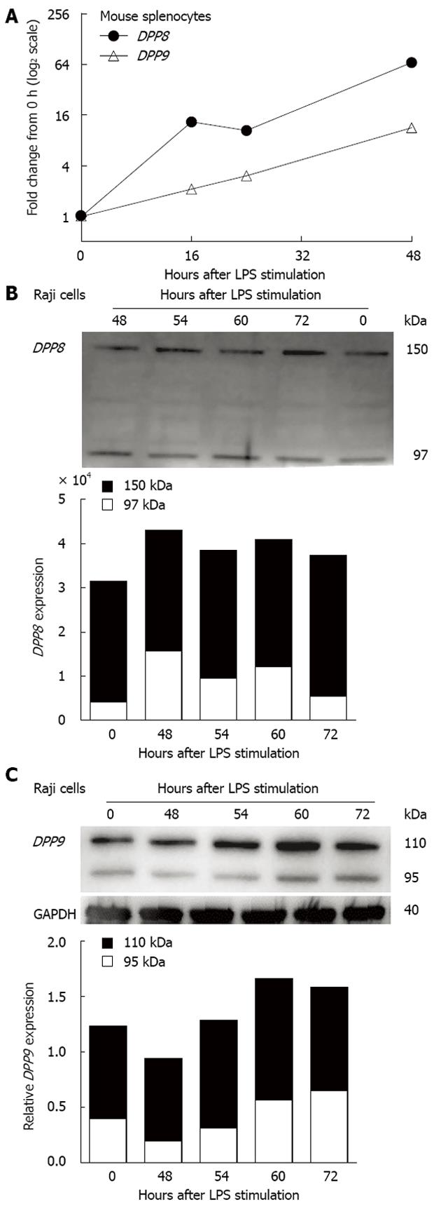
Figure 3 Dipeptidyl peptidase 8 and dipeptidyl peptidase 9 upregulation in lipopolysaccharide stimulated lymphocytes.
A: Dipeptidyl peptidase (DPP) 8 and DPP9 mRNA in mouse splenocytes (representative data from one of three mice); B: Immunoblot of DPP8 and densitometry analysis of DPP8 bands; C: DPP9 immunoblot and densitometry analysis of DPP9 bands relative to glyceraldehyde 3-phosphate dehydrogenase (GAPDH). LPS: Lipopolysaccharide.
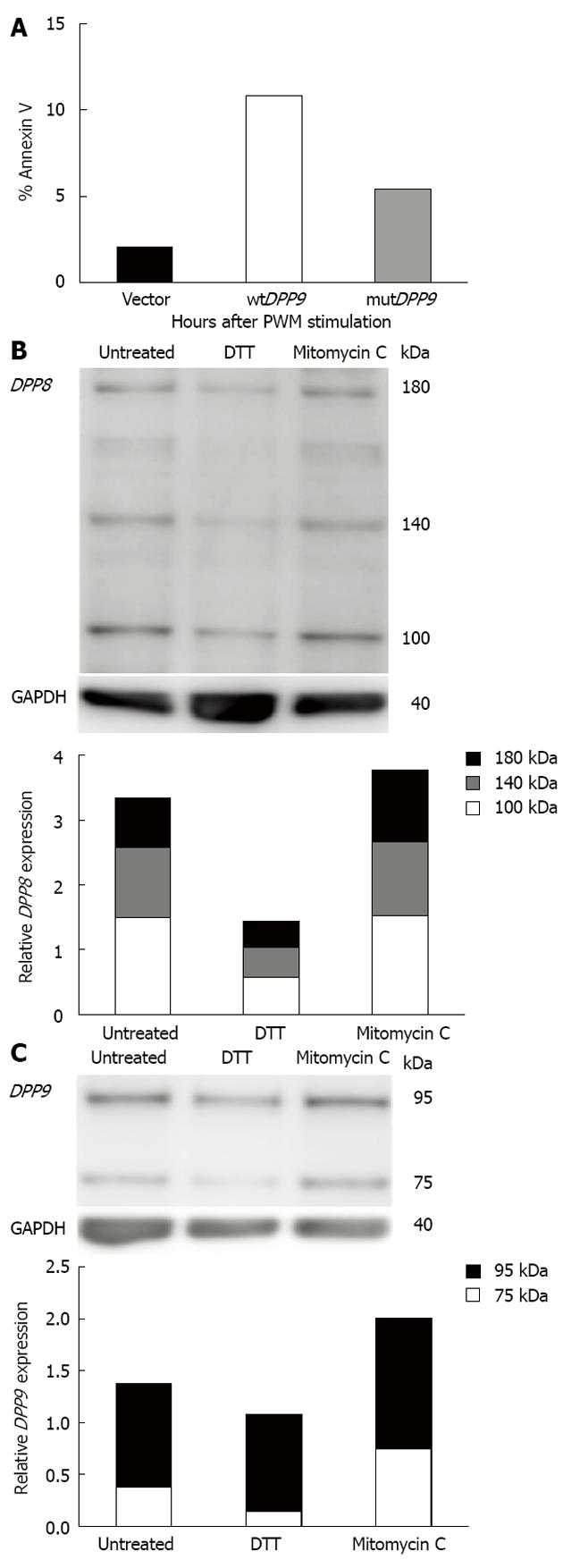
Figure 4 Dipeptidyl peptidase 8 and dipeptidyl peptidase 9 were associated with lymphocyte apoptosis.
A: Percentage of annexin V + Raji cells 40 h after transfection with vector, wild type (wt) dipeptidyl peptidase (DPP) 9-V5-His or enzyme-negative mutant (mut) DPP9-V5-His. Annexin V staining was enumerated by flow cytometry; B: Immunoblot of DPP8 and its densitometry (C) immunoblot of DPP9 and its densitometry in Raji cells untreated and treated with dithiothreitol (DTT) or mitomycin C for 24 h. Densitometry are shown as relative to glyceraldehyde 3-phosphate dehydrogenase (GAPDH).
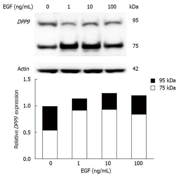
Figure 5 Dipeptidyl peptidase 9 upregulation in epidermal growth factor treated Huh7 cells.
Dipeptidyl peptidase (DPP) 9 immunoblot of untreated and epidermal growth factor (EGF)-treated Huh7 cells at 4 h. Cells were serum starved overnight before EGF treatment. Densitometry of DPP9 is shown relative to actin.
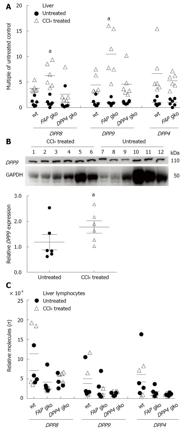
Figure 6 Dipeptidyl peptidase mRNA upregulation in carbon tetrachloride induced liver injury.
A: Multiple of intrahepatic mRNA in carbon tetrachloride (CCl4) treated mice to mean of untreated control mice; aP < 0.05 in CCl4 treated fibroblast activation protein (FAP) gene knockout (gko) vs wild type (wt); B: Dipeptidyl peptidase (DPP) 9 immunoblot of livers from CCl4 treated (lanes 1-6) and untreated mice (lanes 7-12) (n = 6 per group): Densitometry of intrahepatic DPP9 relative to glyceraldehyde 3-phosphate dehydrogenase (GAPDH). aP < 0.05 vs untreated controls; C: mRNA quantitation from isolated hepatic lymphocytes relative to 18S.
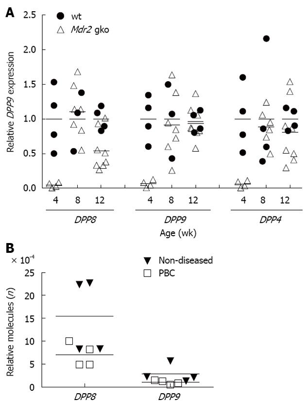
Figure 7 Dipeptidyl peptidase mRNA in mouse and human biliary liver diseases.
A: Multiple of Intrahepatic dipeptidyl peptidase (DPP) mRNA in multidrug resistance gene 2 (Mdr2) gene knockout (gko) female mice to mean of wild type (wt) controls; B: Human end-stage primary biliary cirrhosis (PBC) and non-diseased control livers. Data from each individual is shown as the number of molecules relative to aldolase B (n = 4 per group).
- Citation: Chowdhury S, Chen Y, Yao TW, Ajami K, Wang XM, Popov Y, Schuppan D, Bertolino P, McCaughan GW, Yu DM, Gorrell MD. Regulation of dipeptidyl peptidase 8 and 9 expression in activated lymphocytes and injured liver. World J Gastroenterol 2013; 19(19): 2883-2893
- URL: https://www.wjgnet.com/1007-9327/full/v19/i19/2883.htm
- DOI: https://dx.doi.org/10.3748/wjg.v19.i19.2883


