Copyright
©2013 Baishideng Publishing Group Co.
World J Gastroenterol. Apr 28, 2013; 19(16): 2574-2577
Published online Apr 28, 2013. doi: 10.3748/wjg.v19.i16.2574
Published online Apr 28, 2013. doi: 10.3748/wjg.v19.i16.2574
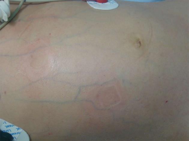
Figure 1 Abdominal bulge was found on examination at admission.
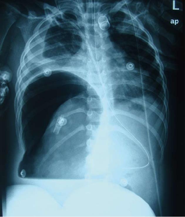
Figure 2 Plain abdominal radiograph in the erect position showed heavy gas in the right subphrenic space with an elevated diaphragm.
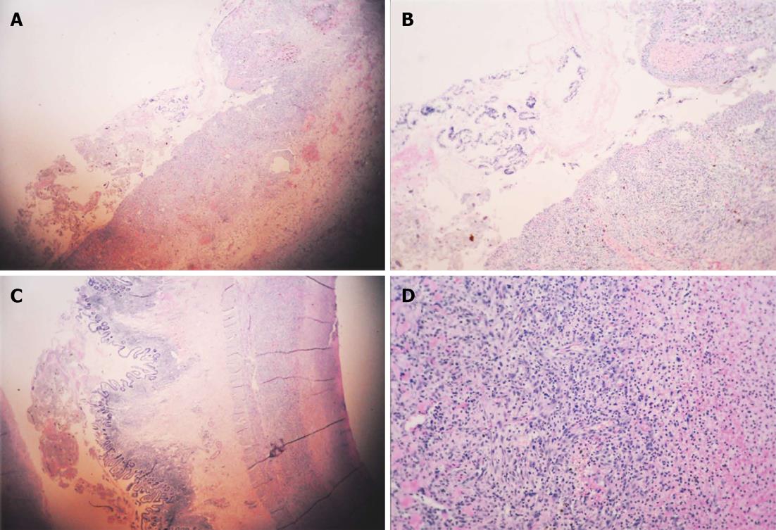
Figure 3 Histopathological analyses of the patient’s ileal biopsy specimens.
A: Hematoxylin and eosin-stained sections of the biopsy specimens revealed mucosal discontinuity (magnification × 10); B: Multiple site bleeding (magnification × 20); C: Inflammatory cell infiltration in all layers of the ileal wall (magnification × 20); D: Infiltration of a large number of neutrophils and cell deformation in the area of edema (magnification × 40).
Figure 4 Cerebral computed tomography demonstrated subarachnoid hemorrhage, right thalamic and intraventricular hemorrhage, and multiple ischemic changes in the left cerebellar hemisphere and bilateral cerebral hemispheres.
A: Right thalamic hemorrhage ruptured into the ventricle; B-D: Thalamic hemorrhage complicated with subarachnoid hemorrhage, cerebral hemispheres multiple focal ischemia.
- Citation: Wang HL, Liu HT, Chen Q, Gao Y, Yu KJ. Henoch-Schonlein purpura with intestinal perforation and cerebral hemorrhage: A case report. World J Gastroenterol 2013; 19(16): 2574-2577
- URL: https://www.wjgnet.com/1007-9327/full/v19/i16/2574.htm
- DOI: https://dx.doi.org/10.3748/wjg.v19.i16.2574


