Copyright
©2013 Baishideng Publishing Group Co.
World J Gastroenterol. Apr 28, 2013; 19(16): 2569-2573
Published online Apr 28, 2013. doi: 10.3748/wjg.v19.i16.2569
Published online Apr 28, 2013. doi: 10.3748/wjg.v19.i16.2569
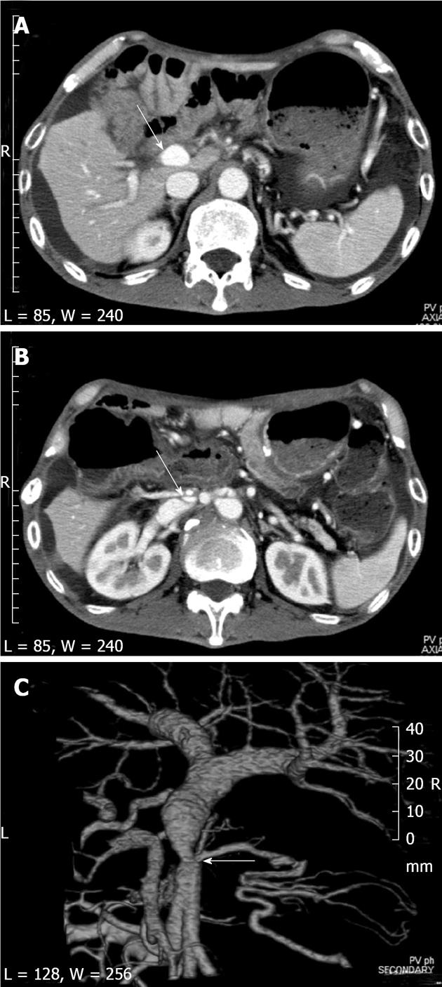
Figure 1 Computed tomography showed short segmental stenosis of the portal vein in the region of the anastomosis, severe ascites, and liver atrophy.
A, B: Computed tomography (CT) scan shows severe ascites and liver atrophy (arrow) (A), and stenosis of the portal vein (arrow) behind the superior mesenteric artery (B); C: The image of the 3D reconstruction of the portal vein shows short segmental stenosis in the region of the anastomosis (arrow).
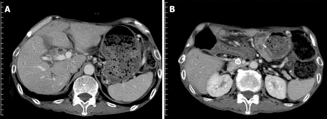
Figure 2 Computed tomography scan 3 mo after the expandable metallic stent placement.
A: Shows that the ascites decreased and the liver atrophy improved; B: The stent placed in the portal vein remained patent.
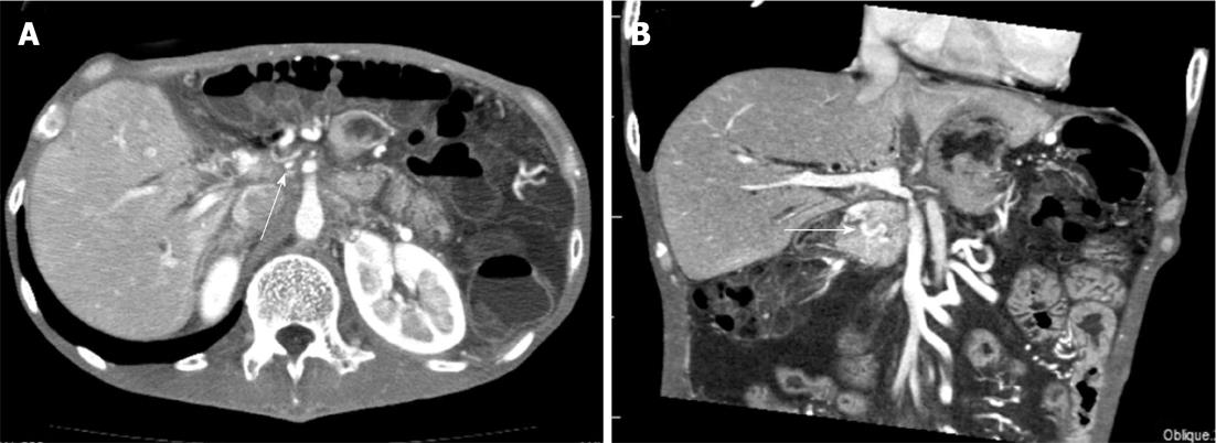
Figure 3 Computed tomography showed short segmental stenosis of the portal vein in the region of the anastomosis, collateral circulation through the cavernous transformation of the pancreatic head, severe ascites, and thickness of the intestinal wall.
A: Computed tomography scan showing severe portal vein stenosis (arrow) in the region of the anastomosis; B: Multiplanar reconstruction revealed the collateral circulation through the cavernous transformation of the pancreatic head (arrow), severe ascites and thickness of the intestinal wall.
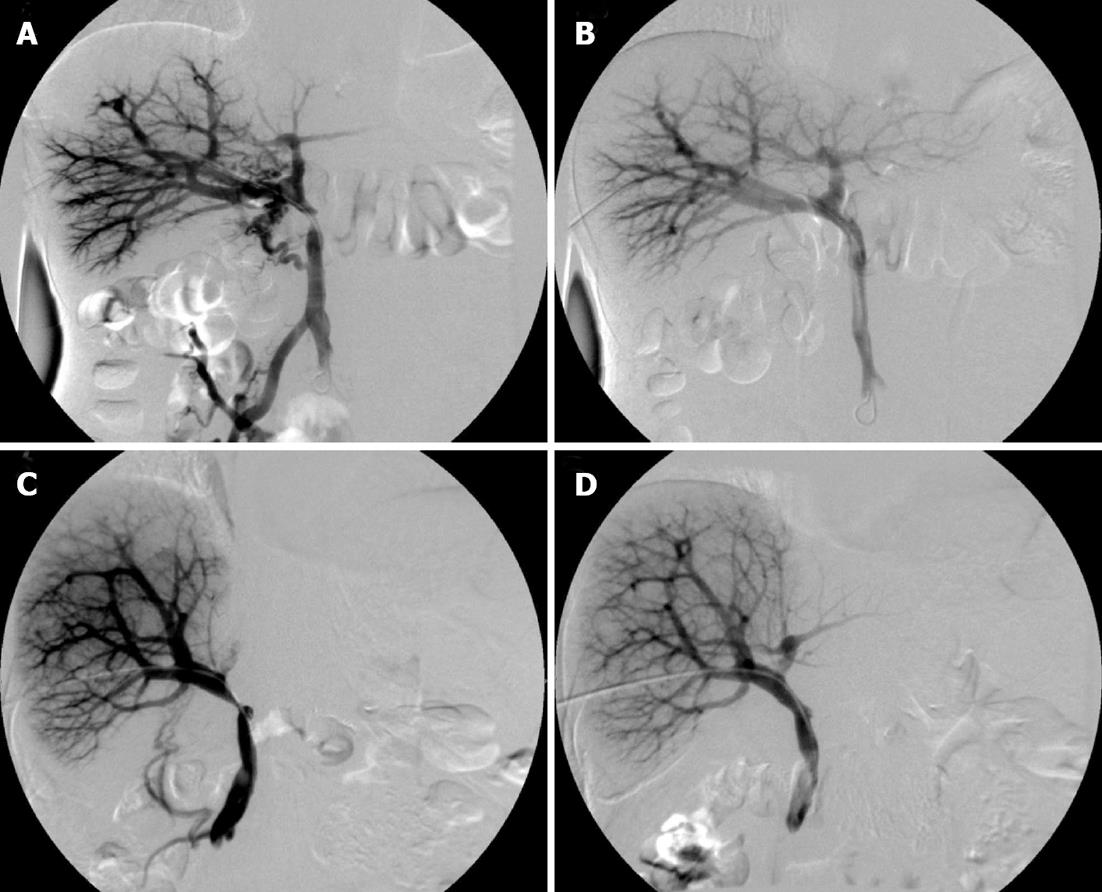
Figure 4 Percutaneous transhepatic direct portography showing short segmental stenosis of the portal vein in the region of the anastomosis.
A: Collateral circulation of the pancreatic head; B: After the expandable metallic stent (EMS) placement, the stenosis was improved, and the collateral circulation disappeared; C: The blood flow of the umbilical portion of the left portal vein was unclear; D: The stenosis was improved, and the blood flow of the umbilical portion became clear after the EMS placement.
- Citation: Tsuruga Y, Kamachi H, Wakayama K, Kakisaka T, Yokoo H, Kamiyama T, Taketomi A. Portal vein stenosis after pancreatectomy following neoadjuvant chemoradiation therapy for pancreatic cancer. World J Gastroenterol 2013; 19(16): 2569-2573
- URL: https://www.wjgnet.com/1007-9327/full/v19/i16/2569.htm
- DOI: https://dx.doi.org/10.3748/wjg.v19.i16.2569


