Copyright
©2013 Baishideng Publishing Group Co.
World J Gastroenterol. Mar 28, 2013; 19(12): 1953-1961
Published online Mar 28, 2013. doi: 10.3748/wjg.v19.i12.1953
Published online Mar 28, 2013. doi: 10.3748/wjg.v19.i12.1953
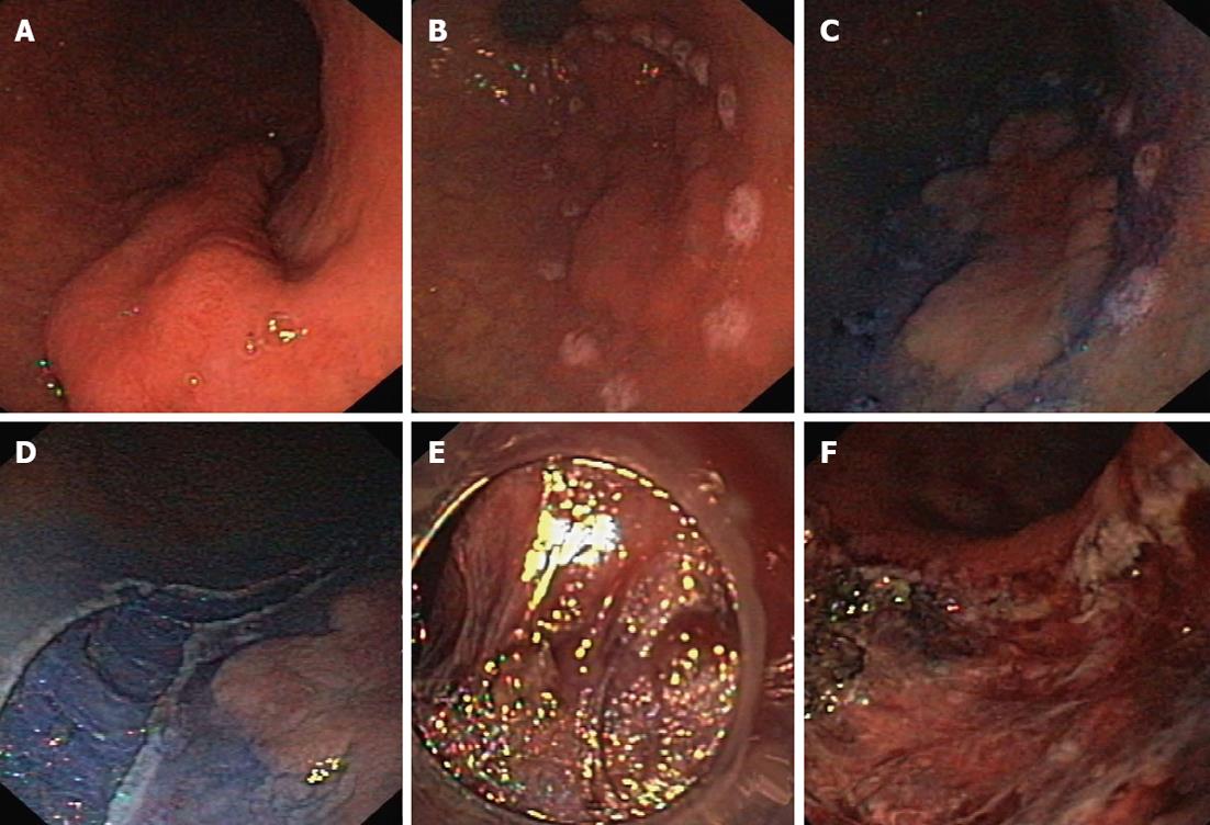
Figure 1 Endoscopic submucosal dissection of gastric neoplastic lesion.
A: Lesion type IIa+c localized in the antrum; B: Margins of the lesion border were marked with the needle knife; C: Solution of indigo carmine in saline was injected into the submucosal space; D: An incision was made in the mucosa and submucosa around the lesion with normal mucosal margin; E: Submucosal dissection performed directly under vision control; F: Mucosal defect after the completed procedure.
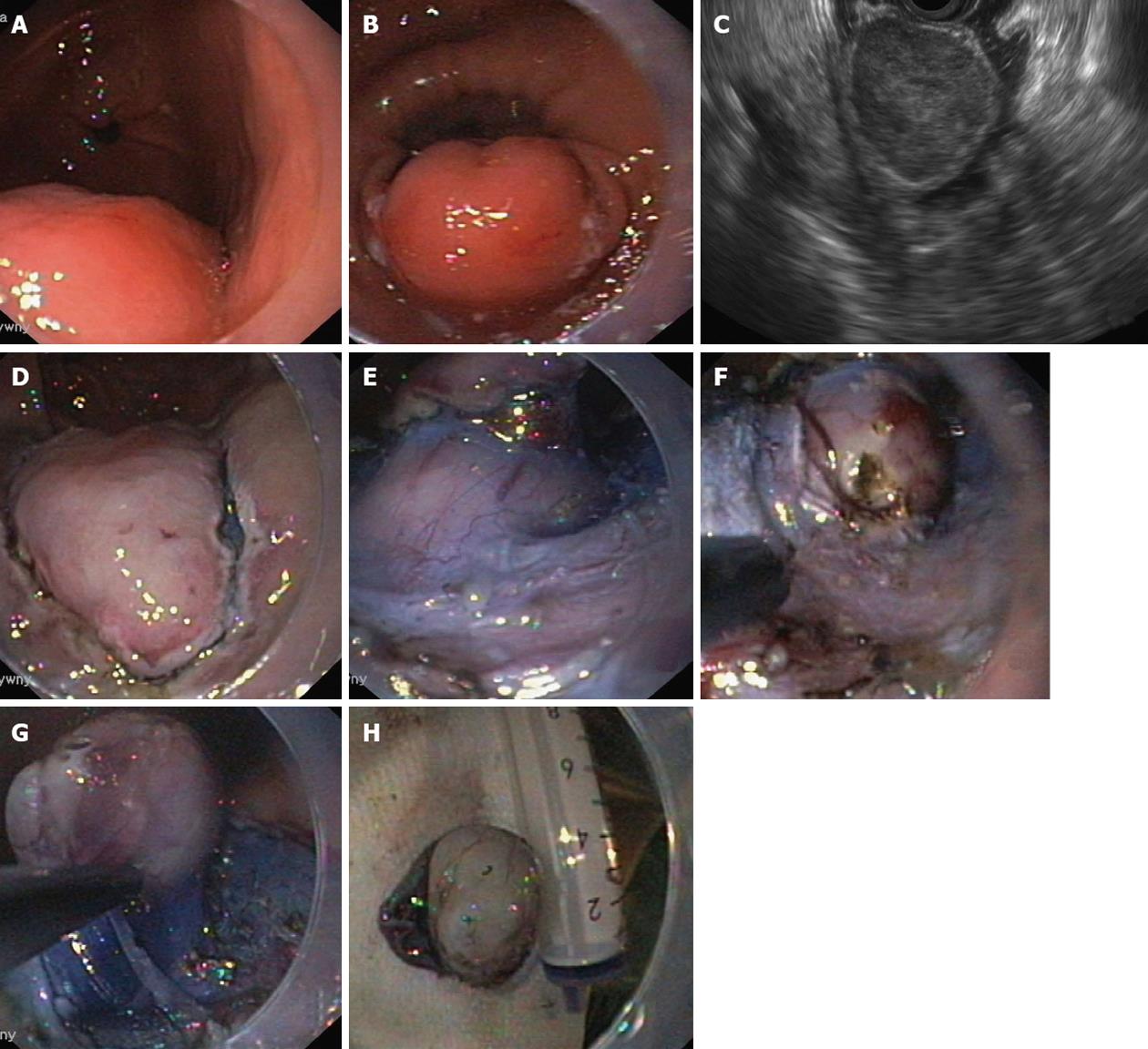
Figure 2 Endoscopic submucosal dissection of gastric submucosal tumor.
A, B: Submucosal tumor in the stomach; C: Endosonographic view of the tumor, which is not connected to the muscle layer of the gastric wall; D: An incision was made in the mucosa and submucosa after indigo carmine solution injection into the submucosal layer around the lesion; E-G: Submucosal dissection of the tumor, exposing the tumor in the submucosa and carefully cutting after injecting the solution; H: Resected tumor (GIST, 25 mm, MI 2/50 HPF). GIST: Gastrointestinal stromal tumor; MI: Mitotic index; HPF: High power field.
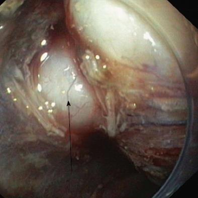
Figure 3 Submucosal tumor (leiomyoma) resected by endoscopic submucosal dissection and connected to the gastric wall with the muscle peduncle (arrow).
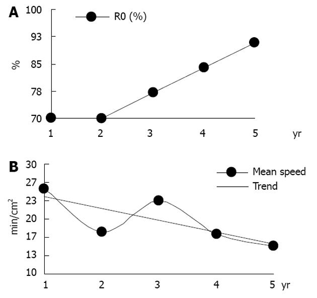
Figure 4 R0 resection rate and mean speed of the procedure in the years following endoscopic submucosal dissection.
A: R0 resection rate; B: Mean speed of the procedure.
- Citation: Białek A, Wiechowska-Kozłowska A, Pertkiewicz J, Karpińska K, Marlicz W, Milkiewicz P, Starzyńska T. Endoscopic submucosal dissection for the treatment of neoplastic lesions in the gastrointestinal tract. World J Gastroenterol 2013; 19(12): 1953-1961
- URL: https://www.wjgnet.com/1007-9327/full/v19/i12/1953.htm
- DOI: https://dx.doi.org/10.3748/wjg.v19.i12.1953


