Copyright
©2013 Baishideng Publishing Group Co.
World J Gastroenterol. Jan 7, 2013; 19(1): 103-107
Published online Jan 7, 2013. doi: 10.3748/wjg.v19.i1.103
Published online Jan 7, 2013. doi: 10.3748/wjg.v19.i1.103
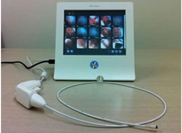
Figure 1 EG scan device.
The EG scan (version I) is composed of a disposable optical probe, a handle, and a display monitor.
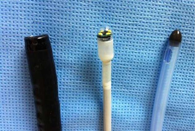
Figure 2 Comparison of diameters.
A: The diameters of GIF-Q260 (Olympus) endoscopy; B: An optical probe of an EG scan device; C: An optical probe of a 16-Fr nasogastric tube.
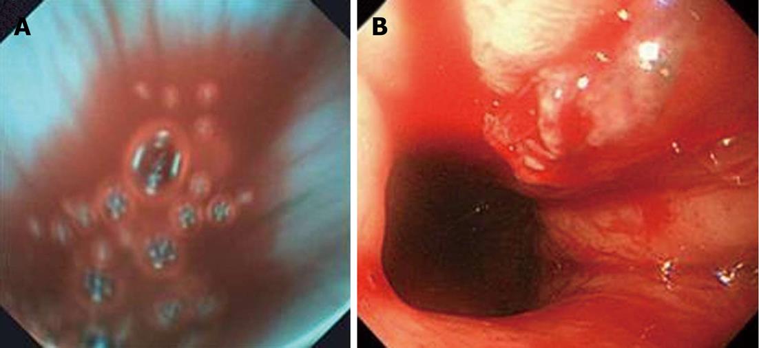
Figure 3 A concordant case of acute gastrointestinal bleeding from Mallory-Weiss syndrome.
A: EG scan shows blood at the esophagogastric junction; B: Conventional esophagogastroduodenoscopy (GIF-Q260) shows oozing from the tear wound of the esophagogastric junction.
- Citation: Cho JH, Kim HM, Lee S, Kim YJ, Han KJ, Cho HG, Song SY. A pilot study of single-use endoscopy in screening acute gastrointestinal bleeding. World J Gastroenterol 2013; 19(1): 103-107
- URL: https://www.wjgnet.com/1007-9327/full/v19/i1/103.htm
- DOI: https://dx.doi.org/10.3748/wjg.v19.i1.103


