Copyright
©2012 Baishideng Publishing Group Co.
World J Gastroenterol. Feb 21, 2012; 18(7): 685-691
Published online Feb 21, 2012. doi: 10.3748/wjg.v18.i7.685
Published online Feb 21, 2012. doi: 10.3748/wjg.v18.i7.685
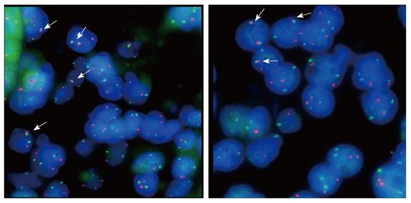
Figure 1 Fluorescence in situ hybridization to detect the translocation (11;18)(q21;121) using the probe LSI API2-MALT1 Dual Color (Vysis, Downers Grove, IL, United States).
Arrows show translocation-positive mucosa-associated lymphoid tissue lymphomas.
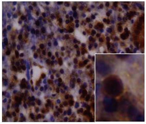
Figure 2 Aberrant expression of B-cell lymphoma 10 protein in gastric mucosa-associated lymphoid tissue lymphoma.
Original magnification, × 400; inset in panel, × 1000.
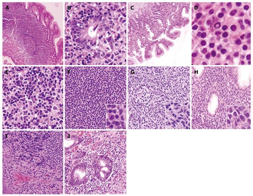
Figure 3 Stain showing tumor morphology (hematoxylin and eosin).
A: Gastric mucosa-associated lymphoid tissue (MALT) lymphoma overview; B: Lymphoepithelial lesion; C: Accompanying intestinal metaplasia in the epithelial tissue surrounding the lymphoma; D: Dutcher’s bodies; E: MALT lymphoma with clusters of large cells; F: Tumor cells of the centrocyte-like/small lymphocyte type; G: Monocytoid tumor cell type; H: Plasma cell differentiation; I: Mixed phenotype composed of centrocytes and plasma cell type; J: Presence of eosinophils. Original magnifications of panels A-J and inset in panel F-H are × 400 and × 1000, respectively.
-
Citation: Yepes S, Torres MM, Saavedra C, Andrade R. Gastric mucosa-associated lymphoid tissue lymphomas and
Helicobacter pylori infection: A Colombian perspective. World J Gastroenterol 2012; 18(7): 685-691 - URL: https://www.wjgnet.com/1007-9327/full/v18/i7/685.htm
- DOI: https://dx.doi.org/10.3748/wjg.v18.i7.685


