Copyright
©2012 Baishideng Publishing Group Co.
World J Gastroenterol. Feb 21, 2012; 18(7): 637-645
Published online Feb 21, 2012. doi: 10.3748/wjg.v18.i7.637
Published online Feb 21, 2012. doi: 10.3748/wjg.v18.i7.637
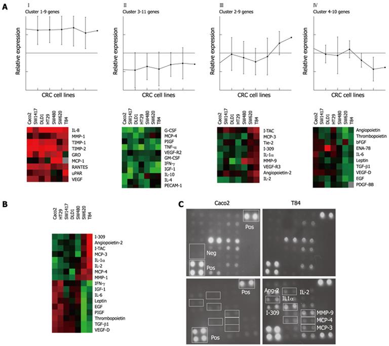
Figure 1 Angiogenesis-related factors expression profile in colorectal cancer cell lines as determined by cytokine antibody-array.
A: K-means (n = 4) clustering grouped the angiogenesis-related proteins according to level of expression; B: Unsupervised-hierarchical clustering of the factors with a significantly different expression in primary and metastatic colorectal cancer (CRC) cell lines; C: Images of subarrays I and II of the primary Caco2 and the metastatic T84 CRC cell lines after detection and processing. IL: Interleukin; MMP: Matrix metalloproteinase; TIMP: Tissue inhibitor of metalloproteinases; GRO: Growth related oncogene; MCP: Macrophage chemoattractant proteins; RANTES: Regulated upon activation normally T-expressed and secreted; uPAR: Urokinase-type plasminogen activator-receptor; G-CSF: Granulocyte colony-stimulating factor; PIGF: Phosphatidylinositol glycan, class F; TNF-α: Tumor necrosis factor-α; GM-CSF: Granulocyte macrophage colony-stimulating factor; IFN-γ: Interferon γ; IGF: Insulin-like growth factor; PECAM: Platelet-endothelial cell adhesion molecule; I-TAC: Inducible T cell α chemoattractant protein; ENA: Epithelial neutrophil activating protein; EGF: Epidermal growth factor; PDGF-BB: Platelet-derived growth factor, β polypeptide; TGF-β1: Transforming growth factor β1; Neg: Negative control; Pos: Positive control.
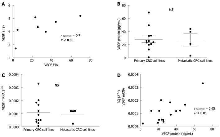
Figure 2 Vascular endothelial growth factor expression in colorectal cancer cell lines.
A: A statistically significant positive correlation is found between vascular endothelial growth factor (VEGF) protein as determined by antibody-array and by enzyme immunoassay (EIA), validating the array method; B and C: Colorectal cancer (CRC) cell lines exhibit variability in VEGF protein (B) and mRNA (C) expression according to their primary or metastatic origin (not statistically significant); D: A statistically significant positive correlation is found between VEGF protein by EIA and VEGF mRNA, suggesting the major role of transcriptional mechanisms in the regulation of VEGF expression. NS: Not significant.
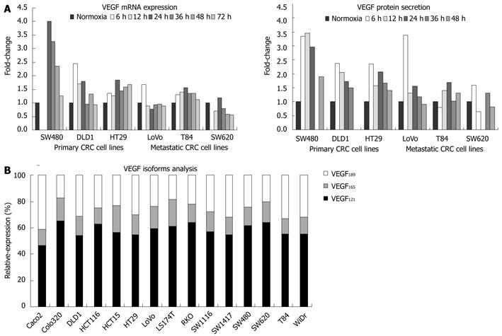
Figure 3 Vascular endothelial growth factor expression regulation.
A: Modulation of vascular endothelial growth factor (VEGF) expression (mRNA and protein) in response to severe hypoxia in primary and metastatic colorectal cancer (CRC) cell lines; B: Expression of VEGF isoforms 121, 189 and 165 by CRC cells in normoxia.
Figure 4 Vascular endothelial growth factor receptors expression in colorectal cancer cell lines.
A: Soluble vascular endothelial growth factor receptor (sVEGFR)-1 expression measured by EIA is not significantly different between primary and metastatic colorectal cancer (CRC) cell lines; B: Flow cytometry of the surface expression of vascular endothelial growth factor receptor (VEGFR)-2 in human umbilical vein endothelial cells (HUVEC) and the primary CRC cell lines HCT116, Caco2 and RKO under normoxic conditions reveals a general lack of VEGFR-2 expression on the surface of CRC cells as compared to HUVEC. NS: Not significant.
- Citation: Abajo A, Bitarte N, Zarate R, Boni V, Lopez I, Gonzalez-Huarriz M, Rodriguez J, Bandres E, Garcia-Foncillas J. Identification of colorectal cancer metastasis markers by an angiogenesis-related cytokine-antibody array. World J Gastroenterol 2012; 18(7): 637-645
- URL: https://www.wjgnet.com/1007-9327/full/v18/i7/637.htm
- DOI: https://dx.doi.org/10.3748/wjg.v18.i7.637


