Copyright
©2012 Baishideng Publishing Group Co.
World J Gastroenterol. Feb 14, 2012; 18(6): 551-556
Published online Feb 14, 2012. doi: 10.3748/wjg.v18.i6.551
Published online Feb 14, 2012. doi: 10.3748/wjg.v18.i6.551
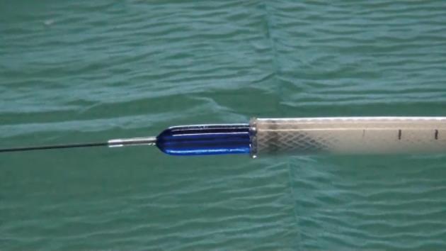
Figure 1 Tracheobronquial Polyflex stent with an esophageal wire-guided balloon inserted through the delivery system with the tip protruding from the introducer by 2 to 3 cm.
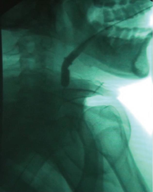
Figure 2 Passage of the stent delivery system through the stricture under fluoroscopic guidance.
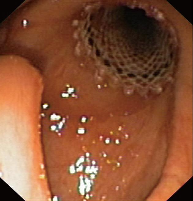
Figure 3 Endoscopic view of the uvula and the stent deployed in the hypopharynx.
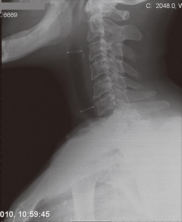
Figure 4 Cervical X-ray to assess adequate positioning and expansion of the stent.
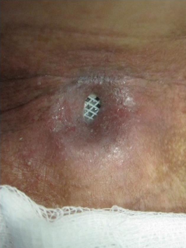
Figure 5 View of the lower end of the stent through the pharyngocutaneous fistula.
- Citation: Silva RA, Mesquita N, Nunes PP, Cardoso E, Pinto RM, Dias LM. Tracheobronchial Polyflex stents for the management of benign refractory hypopharyngeal strictures. World J Gastroenterol 2012; 18(6): 551-556
- URL: https://www.wjgnet.com/1007-9327/full/v18/i6/551.htm
- DOI: https://dx.doi.org/10.3748/wjg.v18.i6.551


