Copyright
©2012 Baishideng Publishing Group Co.
World J Gastroenterol. Feb 7, 2012; 18(5): 401-411
Published online Feb 7, 2012. doi: 10.3748/wjg.v18.i5.401
Published online Feb 7, 2012. doi: 10.3748/wjg.v18.i5.401
Figure 1 Endoscopic images of fundic gastric varices before (A), during (B, C) and after (D) thrombin injection.
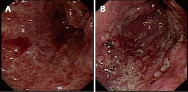
Figure 2 Endoscopic image.
A: Gastric antral vascular ectasia (GAVE) (diffuse type) with active bleeding prior to argon plasma coagulation (APC) treatment; B: GAVE (diffuse type) immediately after APC treatment.
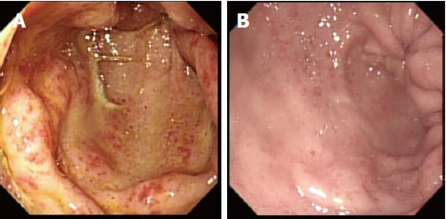
Figure 3 Endoscopic image.
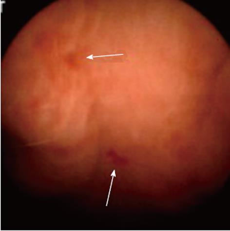
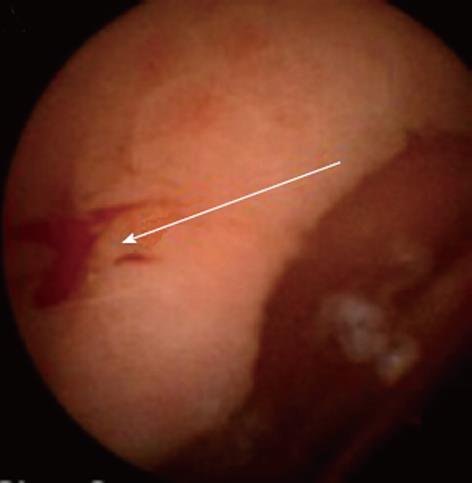
A: Gastric antral vascular ectasia-induced symptomatic anemia; B: Endoscopic image of the same patient 2 years later, after several argon plasma coagulation sessions. The number of angioectatic lesions in the gastric outlet had dramatically decreased.
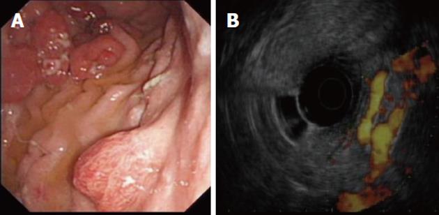
Figure 4 Endoscopic image of gastric irregular submucosal lesion.
A: Gastric irregular submucosal lesion in a patient with portal hypertension; B: The same lesion examined under color Doppler endoscopic ultrasound. The submucosal lesion was hypervascular and represented a gastric varix.
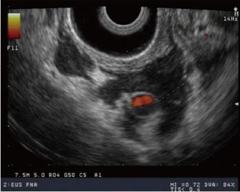
Figure 5 Color doppler endoscopic ultrasound image of duodenal varices after thrombin injection.
The absence of blood flow and the speckled appearances were suggestive of thrombus formation.
Figure 6 Small bowel capsule image of portal hypertensive enteropathy and stigmata of recent bleeding.
Engorged small bowel villi and micro-hemorrhagic spots were visible.
Figure 7 Small bowel capsule image of portal hypertensive enteropathy with snake-skin-like appearance of the mucosa and red spots as stigmata of recent bleeding.
Figure 8 Endomicroscopy image.
A: Image from Cellvizio® bile duct endomicroscopy. The regular reticular pattern of thin dark structures with low signal (dark) characterized the normal bile duct (Image courtesy of http://www. cellvizio.net); B: Abnormal bile duct appearances in Cellvizio® endomicroscopy; isolated blood vessels with very strong signal (with strands) suggestive of tumor neovascularization of cholangiocarcinoma (Image courtesy of http://http://http://www.cellvizio.net); C: Reticular pattern of dark bands and dark clumps or glands suggestive of cholangiocarcinoma (Image courtesy of http://http://http://www.cellvizio.net).
- Citation: Krystallis C, Masterton GS, Hayes PC, Plevris JN. Update of endoscopy in liver disease: More than just treating varices. World J Gastroenterol 2012; 18(5): 401-411
- URL: https://www.wjgnet.com/1007-9327/full/v18/i5/401.htm
- DOI: https://dx.doi.org/10.3748/wjg.v18.i5.401


