Copyright
©2012 Baishideng Publishing Group Co.
World J Gastroenterol. Dec 7, 2012; 18(45): 6686-6689
Published online Dec 7, 2012. doi: 10.3748/wjg.v18.i45.6686
Published online Dec 7, 2012. doi: 10.3748/wjg.v18.i45.6686
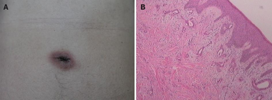
Figure 1 Appearance and pathology of umbilical tumor.
A: A red nodule without ulcer appears in the umbilical region; B: Hematoxylin and eosin stain shows adenocarcinoma cell infiltration (×40).
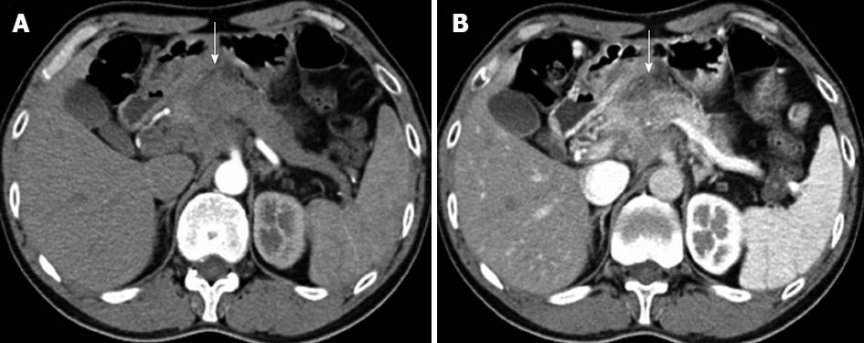
Figure 2 Abdominal contrast-enhanced multi-detector computed tomography scan.
A: A mass in the neck of pancreas (arrow) is shown in arterial phase; B: The mass in venous phase.
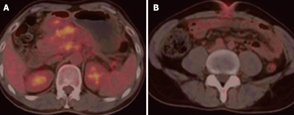
Figure 3 Positron emission tomography-computed tomography showed increased fludeoxyglucose uptake of tumors.
A: Significant enhanced signal showing increased fludeoxyglucose (FDG) uptake at the site of pancreatic mass; B: Increased FDG uptake at the site of umbilical nodule.
- Citation: Bai XL, Zhang Q, Masood W, Masood N, Tang Y, Cao CH, Fu QH, Zhang Y, Gao SL, Liang TB. Sister Mary Joseph’s nodule as a first sign of pancreatic cancer. World J Gastroenterol 2012; 18(45): 6686-6689
- URL: https://www.wjgnet.com/1007-9327/full/v18/i45/6686.htm
- DOI: https://dx.doi.org/10.3748/wjg.v18.i45.6686


