Copyright
©2012 Baishideng Publishing Group Co.
World J Gastroenterol. Dec 7, 2012; 18(45): 6674-6676
Published online Dec 7, 2012. doi: 10.3748/wjg.v18.i45.6674
Published online Dec 7, 2012. doi: 10.3748/wjg.v18.i45.6674
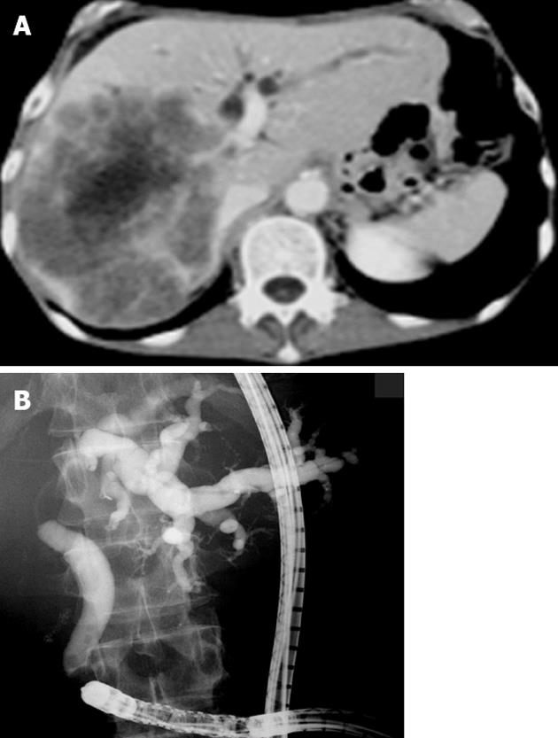
Figure 1 The hilar biliary obstruction due to liver metastasis occupying the right lobe and a dilated left intrahepatic bile duct with tumor invasion extending to the bifurcation of the lateral bile duct branch.
A: Computed tomography image; B: Cholangiography.
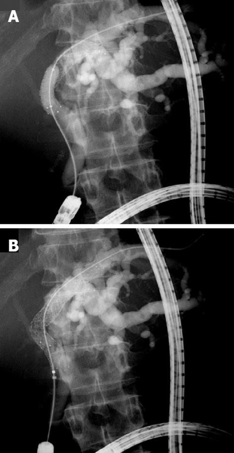
Figure 2 A partial stent-in-stent placement of biliary metallic stents using a short double-balloon enteroscopy.
A: Following the placement of the first self-expandable metallic stent (SEMS), balloon dilation was performed at the stricture; B: The second SEMS was deployed through the mesh of the initial SEMS.
- Citation: Tsutsumi K, Kato H, Tomoda T, Matsumoto K, Sakakihara I, Yamamoto N, Noma Y, Sonoyama T, Okada H, Yamamoto K. Partial stent-in-stent placement of biliary metallic stents using a short double-balloon enteroscopy. World J Gastroenterol 2012; 18(45): 6674-6676
- URL: https://www.wjgnet.com/1007-9327/full/v18/i45/6674.htm
- DOI: https://dx.doi.org/10.3748/wjg.v18.i45.6674


