Copyright
©2012 Baishideng Publishing Group Co.
World J Gastroenterol. Dec 7, 2012; 18(45): 6669-6673
Published online Dec 7, 2012. doi: 10.3748/wjg.v18.i45.6669
Published online Dec 7, 2012. doi: 10.3748/wjg.v18.i45.6669
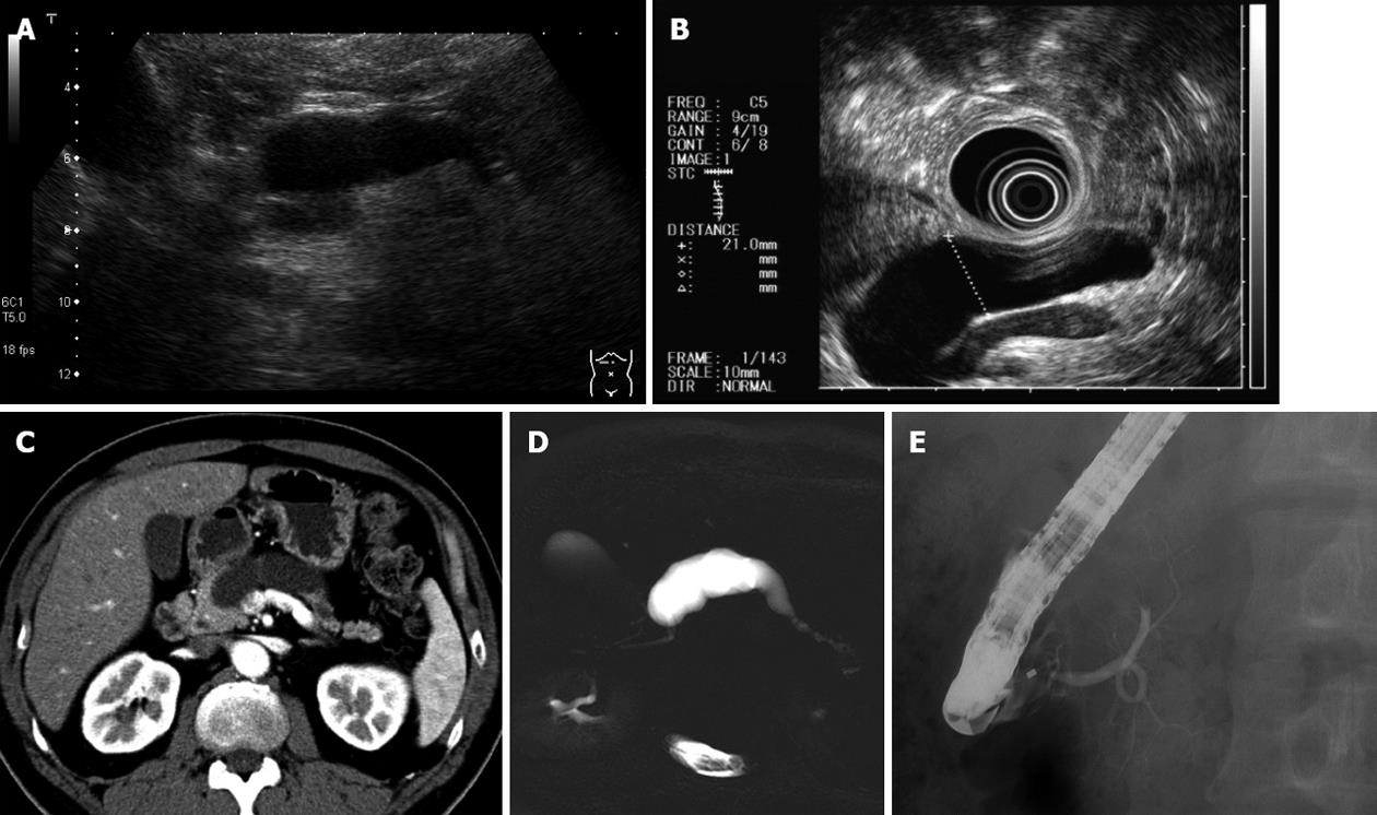
Figure 1 No tumor was detected by endoscopic ultrasonography, computed tomography and magnetic resonance imaging.
The diameter of the main pancreatic duct was > 20 mm at the body. A: Ultrasonography; B: Endoscopic ultrasonography; C: Computed tomography; D: Magnetic resonance cholangiopancreatography; E: Endoscopic retrograde pancreatography.
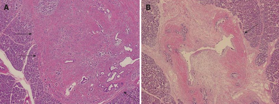
Figure 2 Pathological findings (hematoxylin/eosin staining).
A: A 5 mm × 3 mm tumor was detected (arrows); B: Fibrosis was present around the main pancreatic duct (arrow), and it seemed to cause stricture.
Figure 3 Immunohistochemical staining was positive for chromogranin A, synaptophysin and serotonin.
A: Hematoxylin/eosin staining; B: Chromogranin A; C: Serotonin. Ki-67 labeling index was less than 1%.
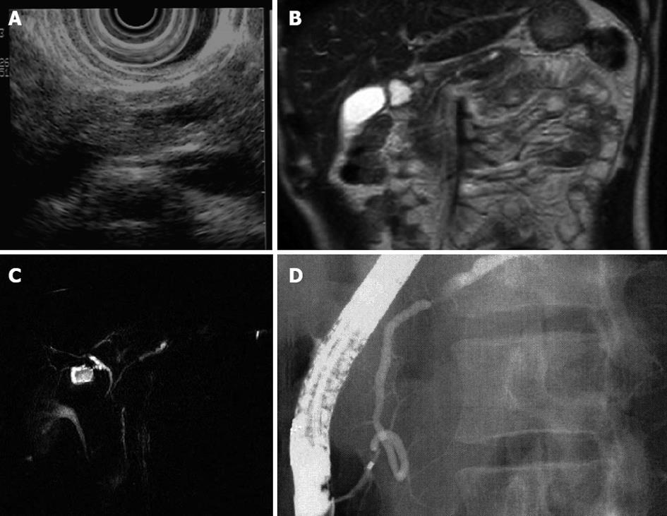
Figure 4 Dilation of the main pancreatic duct.
Endoscopic ultrasonography was demonstrated presence of a small pancreatic tumor. A: Ultrasonography; B: Magnetic resonance imaging; C: Magnetic resonance cholangiopancreatography; D: Endoscopic retrograde pancreatography.
Figure 5 Pathological findings (hematoxylin and eosin staining) and immunohistochemical staining.
A: A 4 mm × 5 mm tumor was detected (arrows); B: Chromogranin A; C: Serotonin. MPD: Main pancreatic duct.
- Citation: Ogawa M, Kawaguchi Y, Maruno A, Ito H, Nakagohri T, Hirabayashi K, Yamamuro H, Yamashita T, Mine T. Small serotonin-positive pancreatic endocrine tumors caused obstruction of the main pancreatic duct. World J Gastroenterol 2012; 18(45): 6669-6673
- URL: https://www.wjgnet.com/1007-9327/full/v18/i45/6669.htm
- DOI: https://dx.doi.org/10.3748/wjg.v18.i45.6669


