Copyright
©2012 Baishideng Publishing Group Co.
World J Gastroenterol. Nov 28, 2012; 18(44): 6501-6503
Published online Nov 28, 2012. doi: 10.3748/wjg.v18.i44.6501
Published online Nov 28, 2012. doi: 10.3748/wjg.v18.i44.6501
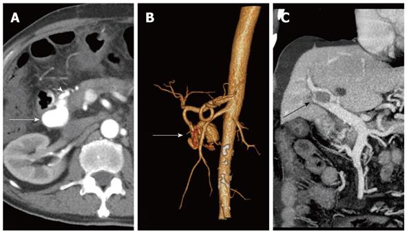
Figure 1 Contrast-enhanced computed tomography scan of the upper abdomen.
A: Computerized tomography (CT) scan revealed a large aneurysm (white arrow) with dilated feeding gastroduodenal artery (GDA) (white arrowhead). Draining superior mesenteric vein (SMV) was observed at a lower level; B: Three-dimensional CT reconstruction demonstrated an arterioportal fistula (white arrow) arising from the GDA; C: CT scan showed a high-density mass in the right portal vein (black arrow), the patent main portal vein and SMV.
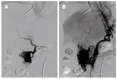
Figure 2 Selective angiography of the gastroduodenal artery.
A: Angiography demonstrated an arterioportal fistula fistulating into the superior mesenteric vein; B: The varices were opacified.
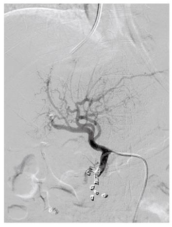
Figure 3 After the placement of several metal coils, the arterioportal fistula was utterly abrogated.
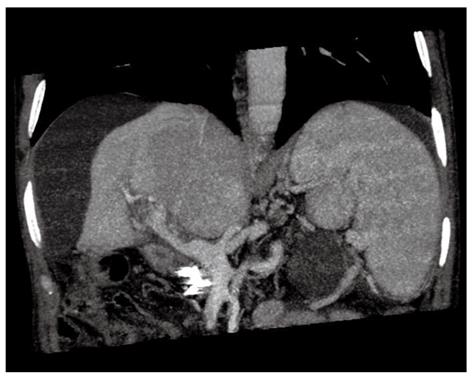
Figure 4 Computerized tomography scan revealed massive intrahepatic portal vein thrombosis and main portal vein thrombosis.
- Citation: Nie L, Luo XF, Li X. Gastrointestinal bleeding caused by extrahepatic arterioportal fistula associated with portal vein thrombosis. World J Gastroenterol 2012; 18(44): 6501-6503
- URL: https://www.wjgnet.com/1007-9327/full/v18/i44/6501.htm
- DOI: https://dx.doi.org/10.3748/wjg.v18.i44.6501


