Copyright
©2012 Baishideng Publishing Group Co.
World J Gastroenterol. Nov 28, 2012; 18(44): 6349-6356
Published online Nov 28, 2012. doi: 10.3748/wjg.v18.i44.6349
Published online Nov 28, 2012. doi: 10.3748/wjg.v18.i44.6349
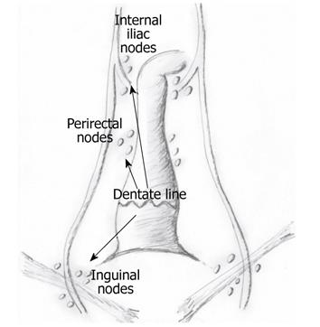
Figure 1 Anal canal lymphatic drainage pattern.
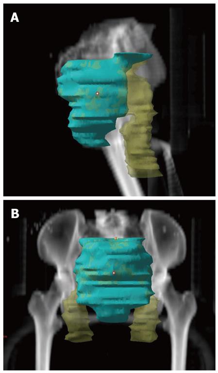
Figure 2 Radiation field of anal carcinoma with exclusion (green field) or inclusion of inguinal regions.
A: Lateral view; B: Anterior view.
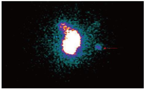
Figure 3 Lymphoscintigraphy of a sentinel lymph node in anal carcinoma.
Anterior view showing injection site (blue arrow) and sentinel lymph node (red arrow).
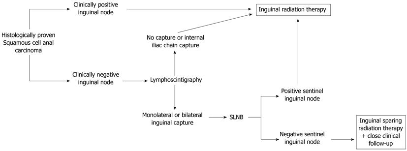
Figure 4 Diagnostic-therapeutic algorithm for squamous cell anal carcinoma.
SLNB: Sentinel lymph node biopsy.
- Citation: De Nardi P, Carvello M, Staudacher C. New approach to anal cancer: Individualized therapy based on sentinel lymph node biopsy. World J Gastroenterol 2012; 18(44): 6349-6356
- URL: https://www.wjgnet.com/1007-9327/full/v18/i44/6349.htm
- DOI: https://dx.doi.org/10.3748/wjg.v18.i44.6349


