Copyright
©2012 Baishideng Publishing Group Co.
World J Gastroenterol. Nov 14, 2012; 18(42): 6155-6159
Published online Nov 14, 2012. doi: 10.3748/wjg.v18.i42.6155
Published online Nov 14, 2012. doi: 10.3748/wjg.v18.i42.6155
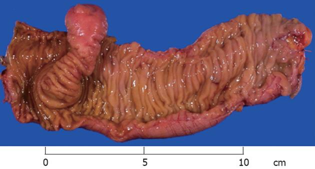
Figure 1 Gross specimen of the inverted Meckel’s diverticulum arising in the segmental resection of the small bowel.
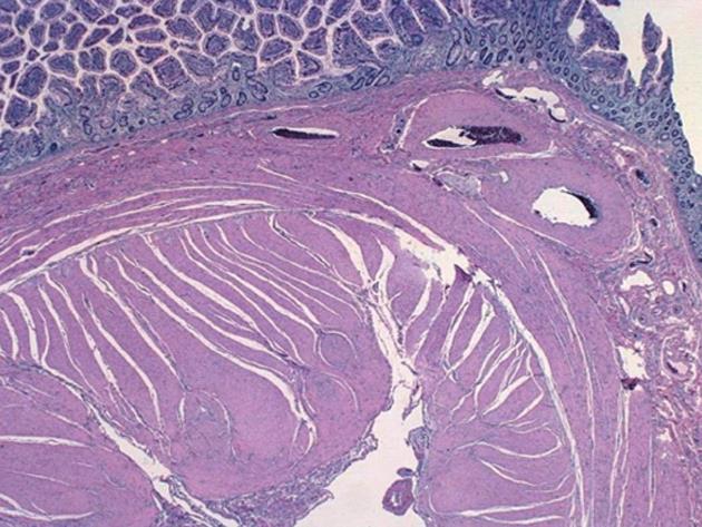
Figure 2 Histopathology reveals a central core consisting of serosa and muscle.
The cross section reveals small intestinal mucosa lining the fragment.
- Citation: Rashid OM, Ku JK, Nagahashi M, Yamada A, Takabe K. Inverted Meckel's diverticulum as a cause of occult lower gastrointestinal hemorrhage. World J Gastroenterol 2012; 18(42): 6155-6159
- URL: https://www.wjgnet.com/1007-9327/full/v18/i42/6155.htm
- DOI: https://dx.doi.org/10.3748/wjg.v18.i42.6155


