Copyright
©2012 Baishideng Publishing Group Co.
World J Gastroenterol. Nov 7, 2012; 18(41): 5965-5971
Published online Nov 7, 2012. doi: 10.3748/wjg.v18.i41.5965
Published online Nov 7, 2012. doi: 10.3748/wjg.v18.i41.5965
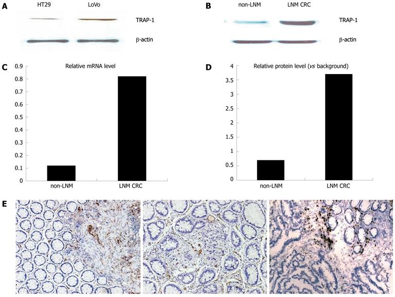
Figure 1 Confirmation of the overexpression of tumor necrosis factor receptor-associated protein-1 in colorectal cancer.
A: Western blotting analysis for tumor necrosis factor receptor-associated protein-1 (TRAP-1) expression in different metastatic potential LoVo cell and HT29 cell. β-actin was used as the internal loading control. The histogram shows the relative expression levels of TRAP-1 in LoVo and HT29 cell; B: Western blotting analysis for TRAP-1 expression in non-lymph node metastasis (LNM) and LNM groups. Data represent the mean ± SE (P < 0.001, Student t test); C, D: mRNA level of TRAP-1 via quantitative real-time polymerase chain reaction. TRAP-1 was consistently increased in the LNM group compared with non-LNM group. The mRNA level was normalized to that of β-actin. Data represent the mean ± SE (P < 0.001, Student t test); E: Immunohistochemical labeling for the TRAP-1 in the CRC sample. TRAP-1 was identified in non-LNM cancer tissues (weak in middle) and strong staining in LNM cancer group (right), but was rare in normal mucosa (left).
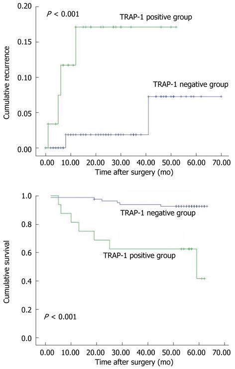
Figure 2 Overexpression of tumor necrosis factor receptor-associated protein-1 correlated with poor prognosis in 96 colorectal cancer patients.
A: Cumulative recurrence; B: Cumulative survival. The tumor necrosis factor receptor-associated protein-1 (TRAP-1)-positive vs TRAP-1-negative groups (P < 0.001, log-rank test).
- Citation: Gao JY, Song BR, Peng JJ, Lu YM. Correlation between mitochondrial TRAP-1 expression and lymph node metastasis in colorectal cancer. World J Gastroenterol 2012; 18(41): 5965-5971
- URL: https://www.wjgnet.com/1007-9327/full/v18/i41/5965.htm
- DOI: https://dx.doi.org/10.3748/wjg.v18.i41.5965


