Copyright
©2012 Baishideng Publishing Group Co.
World J Gastroenterol. Oct 14, 2012; 18(38): 5434-5441
Published online Oct 14, 2012. doi: 10.3748/wjg.v18.i38.5434
Published online Oct 14, 2012. doi: 10.3748/wjg.v18.i38.5434
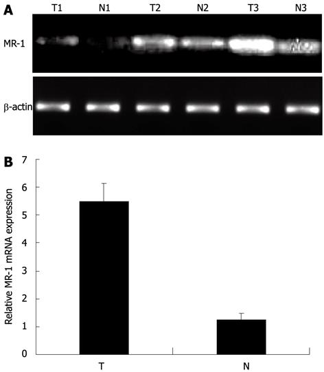
Figure 1 Expression of myofibrillogenesis regulator-1 mRNA in adjacent noncancerous mucosa and gastric cancers.
A: Expression pattern of myofibrillogenesis regulator-1 (MR-1) in gastric cancer specimens by semiquan- titative reverse transcription-polymerase chain reaction (PCR) (representative PCR results from 3 patients). The expression level of MR-1 mRNA showed significant difference between gastric cancer tissues and corresponding noncancerous gastric tissues; B: Results of real-time quantitative PCR assay. MR-1 mRNA expression in tumor tissue was frequently higher than that in matched normal mucosa (P < 0.001; Student’s t test). β-actin was used as a control. N: Noncancerous gastric tissues; T: Gastric cancer tissues.
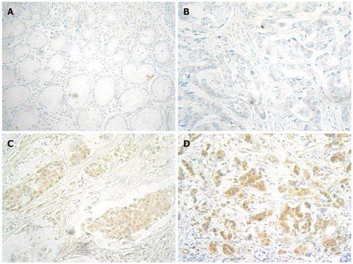
Figure 2 Myofibrillogenesis regulator-1 protein expression determined by immunohistochemical staining in adjacent noncancerous mucosa and gastric cancers.
A: Negative expression in adjacent noncancerous mucosa (× 200). No myofibrillogenesis regulator-1 (MR-1) expression was detected; B: Negative expression in gastric cancers (× 200). No MR-1 expression was detected; C: Weak positive expression in gastric cancer (× 200); D: Moderate positive expression in gastric cancer (× 200). MR-1 protein was expressed in cytoplasm of the cells in (C) and (D).
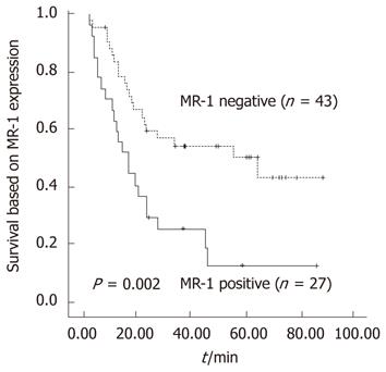
Figure 3 Postoperative survival curves in patients with stage I-IV carcinoma.
Myofibrillogenesis regulator-1 (MR-1) protein positive expression refers to cases showing weak and moderate staining.
- Citation: Guo J, Dong B, Ji JF, Wu AW. Myofibrillogenesis regulator-1 overexpression is associated with poor prognosis of gastric cancer patients. World J Gastroenterol 2012; 18(38): 5434-5441
- URL: https://www.wjgnet.com/1007-9327/full/v18/i38/5434.htm
- DOI: https://dx.doi.org/10.3748/wjg.v18.i38.5434


