Copyright
©2012 Baishideng Publishing Group Co.
World J Gastroenterol. Oct 7, 2012; 18(37): 5305-5308
Published online Oct 7, 2012. doi: 10.3748/wjg.v18.i37.5305
Published online Oct 7, 2012. doi: 10.3748/wjg.v18.i37.5305
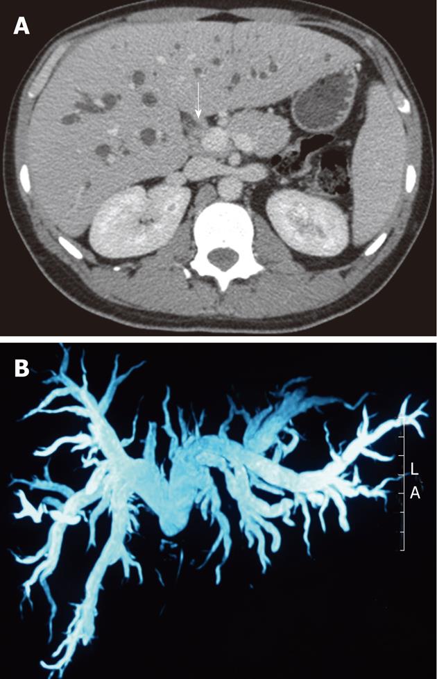
Figure 1 Large dilatation close to hepatic duct.
A: Stenosis and thickening of the common hepatic duct (arrow) with upstream biliary dilation; B: Magnetic resonance cholangiography showing large dilatation close to the lesion in the common hepatic duct. A: Ahead; L: Left.
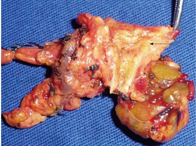
Figure 2 Hepatocholedochal tumor opened longitudinally with the affected area (arrow).
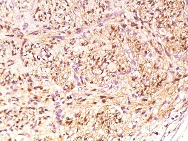
Figure 3 Immunohistochemical staining for the S-100 protein (40 ×).
- Citation: Fonseca GM, Montagnini AL, Rocha MS, Patzina RA, Bernardes MVAA, Cecconello I, Jukemura J. Biliary tract schwannoma: A rare cause of obstructive jaundice in a young patient. World J Gastroenterol 2012; 18(37): 5305-5308
- URL: https://www.wjgnet.com/1007-9327/full/v18/i37/5305.htm
- DOI: https://dx.doi.org/10.3748/wjg.v18.i37.5305


