Copyright
©2012 Baishideng Publishing Group Co.
World J Gastroenterol. Oct 7, 2012; 18(37): 5266-5275
Published online Oct 7, 2012. doi: 10.3748/wjg.v18.i37.5266
Published online Oct 7, 2012. doi: 10.3748/wjg.v18.i37.5266
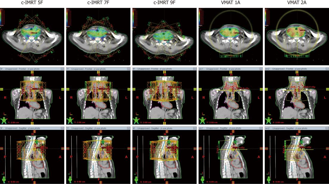
Figure 1 Dose distributions in a cervical esophageal cancer patient planed by conventional sliding window intensity-modulated radiotherapy (5 fields, 7 fields and 9 fields) and volumetric-modulated arc therapy (1 arc and 2 arcs).
IMRT: Intensity-modulated radiotherapy; VMAT: Volumetric modulated arc therapy; F: Coplanar field; A: Arc; Orange line: Planning target volume; Blue line: Spinal cord; Color wash areas: Receiving ≥ 100% of the dose (60 Gy).
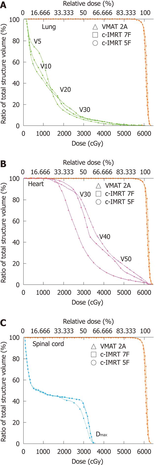
Figure 2 Dose-volume histogram of organs at risk and planning target volume for volumetric-modulated arc therapy and conventional sliding window intensity-modulated radiotherapy in a lower thoracic esophageal cancer patient.
Volumetric-modulated arc therapy (VMAT) with double arcs (triangle) and conventional sliding window intensity-modulated radiotherapy (c-IMRT) with 7 fields (squares) and 5 fields (round). The planning target volume is shown in orange, the lungs in green (A), heart in red (B) and spinal cord in blue (C). F: Coplanar field; A: Arc; VX: The percentage of organ receiving a dose > X Gy.
-
Citation: Yin L, Wu H, Gong J, Geng JH, Jiang F, Shi AH, Yu R, Li YH, Han SK, Xu B, Zhu GY. Volumetric-modulated arc therapy
vs c-IMRT in esophageal cancer: A treatment planning comparison. World J Gastroenterol 2012; 18(37): 5266-5275 - URL: https://www.wjgnet.com/1007-9327/full/v18/i37/5266.htm
- DOI: https://dx.doi.org/10.3748/wjg.v18.i37.5266


