Copyright
©2012 Baishideng Publishing Group Co.
World J Gastroenterol. Oct 7, 2012; 18(37): 5231-5239
Published online Oct 7, 2012. doi: 10.3748/wjg.v18.i37.5231
Published online Oct 7, 2012. doi: 10.3748/wjg.v18.i37.5231
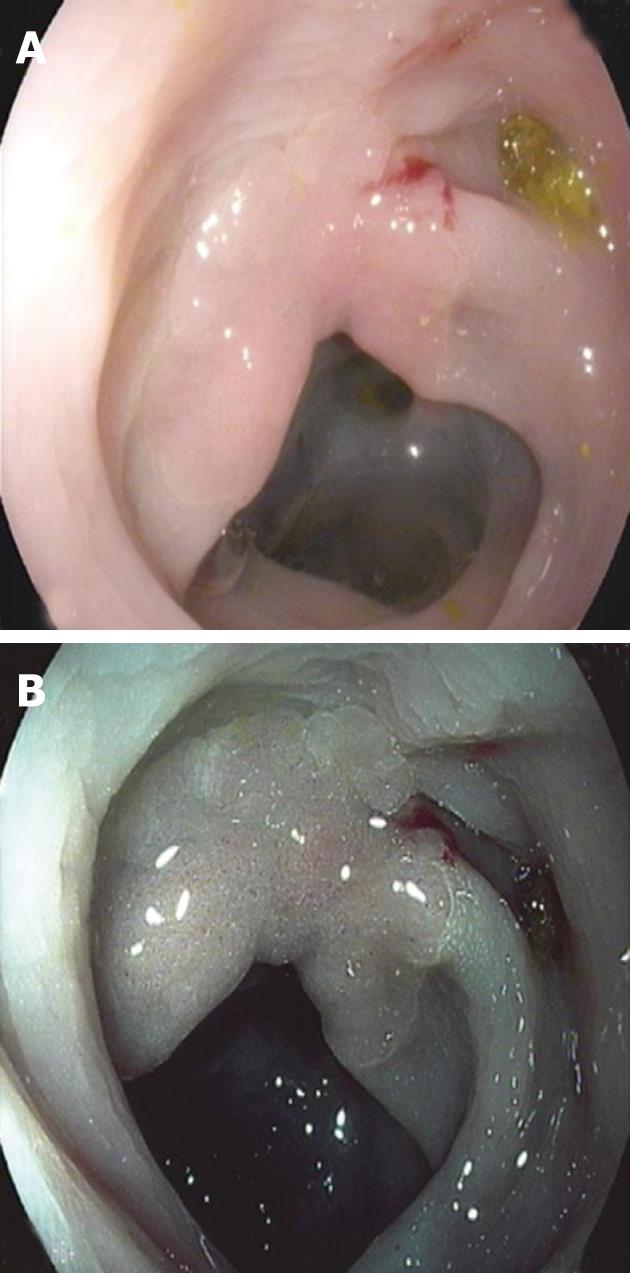
Figure 1 Flat lesion IIb + IIa on left colon examinated by high-definition-white light and visualized with i-Scan.
A: Flat lesion IIb + IIa of 25 mm × 25 mm on left colon examinated by high-definition white light; B: Same lesion visualized with i-Scan.
Figure 2 Flat lesion 0-IIa visualized with high-definition white light and surface enhancement and visualized with i-Scan and digital chromoendoscopy.
A: Flat lesion 0-IIa visualized with high-definition white light and surface enhancement; B: Same lesion visualized with i-Scan and digital chromoendoscopy.
- Citation: Testoni PA, Notaristefano C, Vailati C, Leo MD, Viale E. High-definition colonoscopy with i-Scan: Better diagnosis for small polyps and flat adenomas. World J Gastroenterol 2012; 18(37): 5231-5239
- URL: https://www.wjgnet.com/1007-9327/full/v18/i37/5231.htm
- DOI: https://dx.doi.org/10.3748/wjg.v18.i37.5231


