Copyright
©2012 Baishideng Publishing Group Co.
World J Gastroenterol. Oct 7, 2012; 18(37): 5205-5210
Published online Oct 7, 2012. doi: 10.3748/wjg.v18.i37.5205
Published online Oct 7, 2012. doi: 10.3748/wjg.v18.i37.5205
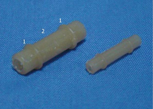
Figure 1 Biodegradable stents of different sizes.
1: Hump rings; 2: Main spindle, 10 mm in length between two hump rings.
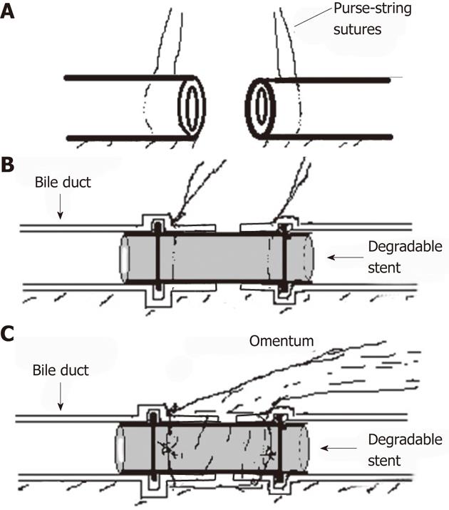
Figure 2 Schematic diagram of repairing bile duct defect with degradable stent and omentum.
A: Purse-string sutures with 4-0 Vicryl were made on both ends of bile duct; B: The stent was inserted into both ends of bile duct; C: A vascularized greater omentum was placed around the stent and both ends of common bile duct.
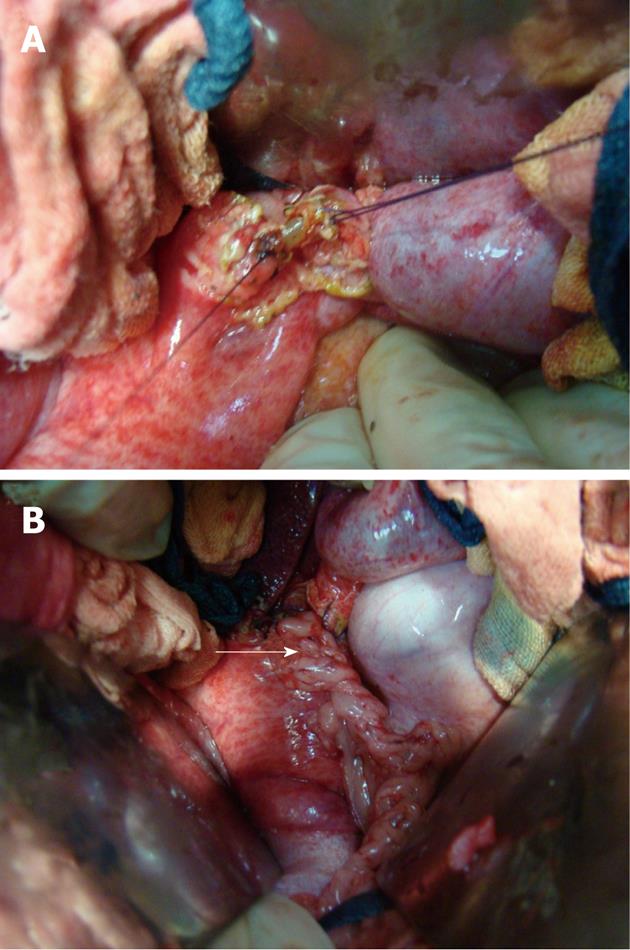
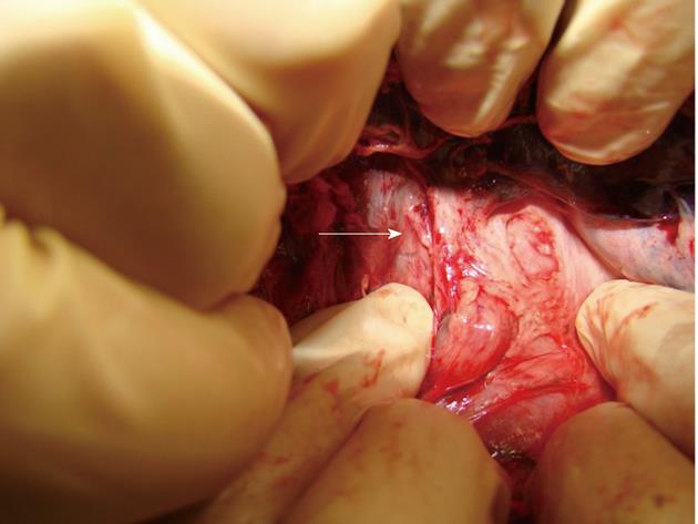
Figure 3 Surgical procedure.
A: Stent is placed into common bile duct and two purse-string sutures are tied; B: A layer of large omentum is covered around the stent and two ends of bile duct (arrow).
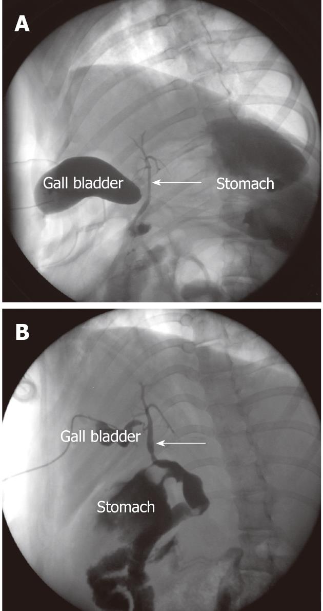
Figure 4 Cholangiography through gall bladder.
A: One month after operation, the shape of the stent (arrow) could be visualized in common bile duct; B: Three months after operation, the stent disappeared.
Figure 5 The common bile duct (arrow) three months after operation.
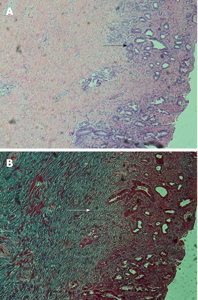
Figure 6 Histology of anastomosis.
A: Hematoxylin and eosin stain × 100, black arrow indicates proliferation of bile duct glands; B: Masson’s trichrome stain × 100, white arrow indicates deposited collagen fibers.
- Citation: Liang YL, Yu YC, Liu K, Wang WJ, Ying JB, Wang YF, Cai XJ. Repair of bile duct defect with degradable stent and autologous tissue in a porcine model. World J Gastroenterol 2012; 18(37): 5205-5210
- URL: https://www.wjgnet.com/1007-9327/full/v18/i37/5205.htm
- DOI: https://dx.doi.org/10.3748/wjg.v18.i37.5205


