Copyright
©2012 Baishideng Publishing Group Co.
World J Gastroenterol. Sep 28, 2012; 18(36): 5135-5137
Published online Sep 28, 2012. doi: 10.3748/wjg.v18.i36.5135
Published online Sep 28, 2012. doi: 10.3748/wjg.v18.i36.5135
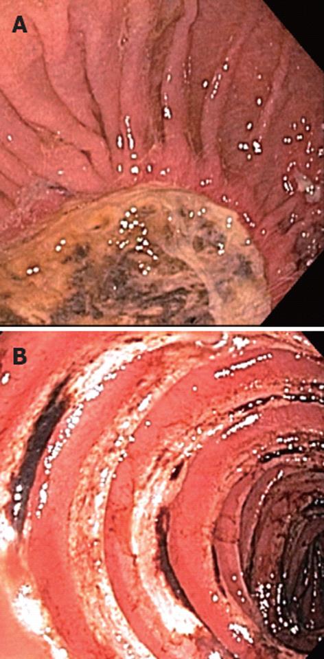
Figure 1 Enteroscopic appearance of the lesions.
A: A 4-cm chronic cavity consistent with chronic perforation at the jejunal anastomosis; B: More distal jejunal ulcerations.
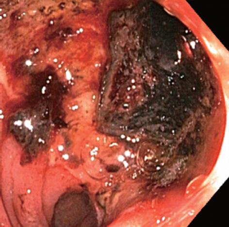
Figure 2 Endoscopy showing bleeding from an anastomotic ulcer.
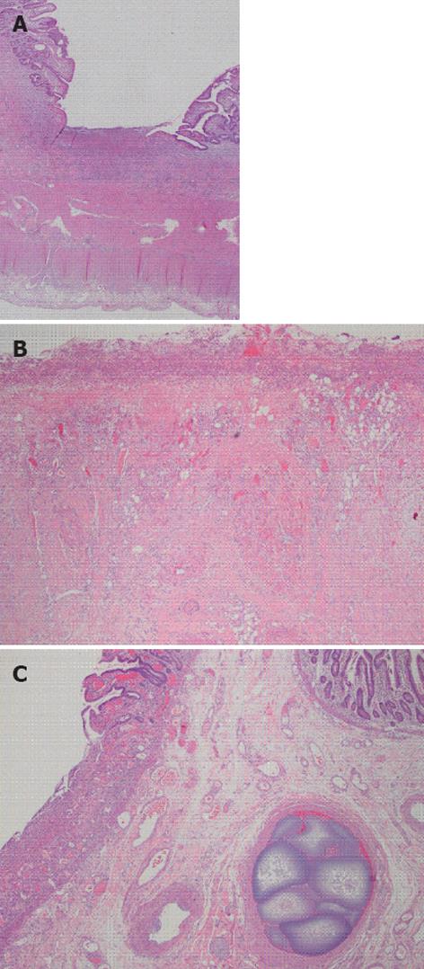
Figure 3 Surgically resected jejunum.
A: Full thickness small bowel section showing ulcerated small intestinal mucosa; B: Ulcer bed showing acute and chronic inflammation, granulation tissue and overlying fibrinopurulent exudate, no granulomas are seen; C: Intravascular embolization material in submucosal blood vessel.
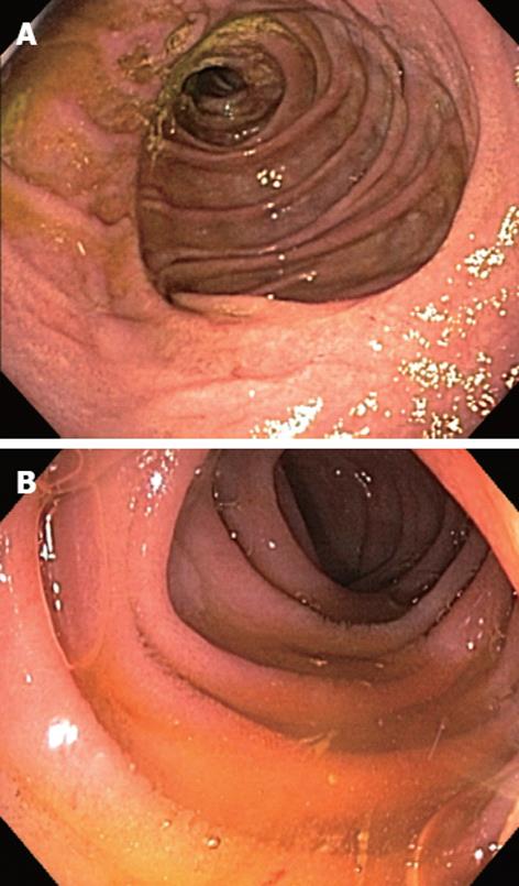
Figure 4 Complete improvement on endoscopic findings after infliximab therapy.
A: Jejunojejunostomy; B: Distal jejunum.
- Citation: Seven G, Assaad A, Biehl T, Kozarek RA. Use of anti tumor necrosis factor-alpha monoclonal antibody for ulcerative jejunoileitis. World J Gastroenterol 2012; 18(36): 5135-5137
- URL: https://www.wjgnet.com/1007-9327/full/v18/i36/5135.htm
- DOI: https://dx.doi.org/10.3748/wjg.v18.i36.5135


