Copyright
©2012 Baishideng Publishing Group Co.
World J Gastroenterol. Sep 28, 2012; 18(36): 5034-5041
Published online Sep 28, 2012. doi: 10.3748/wjg.v18.i36.5034
Published online Sep 28, 2012. doi: 10.3748/wjg.v18.i36.5034
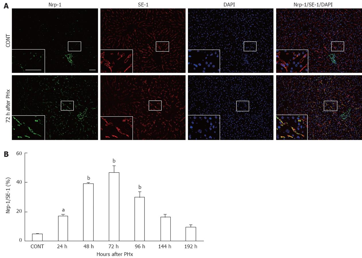
Figure 1 Neuropilin-1 expression in sinusoidal endothelial cells in regenerating liver.
A: Immunofluorescent examination of neuropilin-1 (Nrp-1) in control liver and regenerating liver 72 h after partial hepatectomy. Green: Nrp-1; Red: Sinusoidal endothelial cells stained with SE-1; Blue: Nuclei stained with 4,6-diamidino-2-phenylindole dihydrochloride (DAPI). Scale bar, 50 μm; B: Graphic representation of the percentage of immunostained area with anti-Nrp-1 antibody in relation to the area stained with SE-1 (%). Results are expressed as the mean ± SE. aP < 0.05, bP < 0.01 vs control liver (n = 3). PHx: Partial hepatectomy.
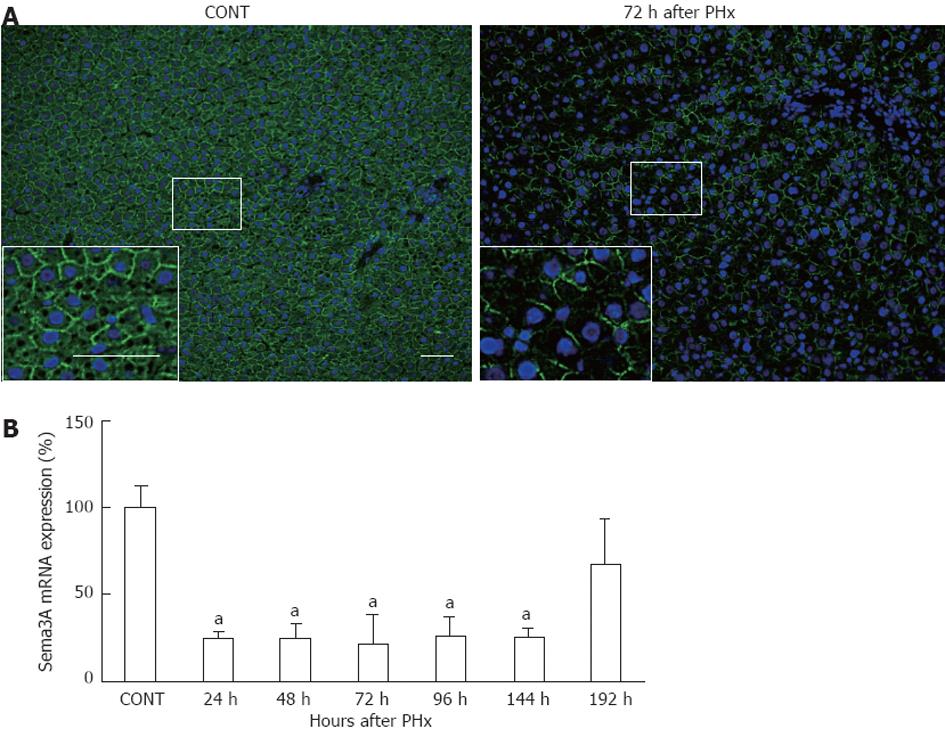
Figure 2 Semaphorin 3A expression in liver tissues after partial hepatectomy.
A: Immunofluorescent examination of semaphorin 3A (Sema3A) in control liver and regenerating liver 72 h after partial hepatectomy (PHx). Green: Sema3A; Blue: Nuclei stained with 4,6-dia-midino-2-phenylindole dihydrochloride. Scale bar, 50 μm; B: Sema3A mRNA expression in liver tissues after PHx. Sema3A mRNA expression was quantified by real-time polymerase chain reaction and normalized to ribosomal protein S18 mRNA level. Results are expressed as the mean ± SE. aP < 0.05 vs control liver (n = 3).
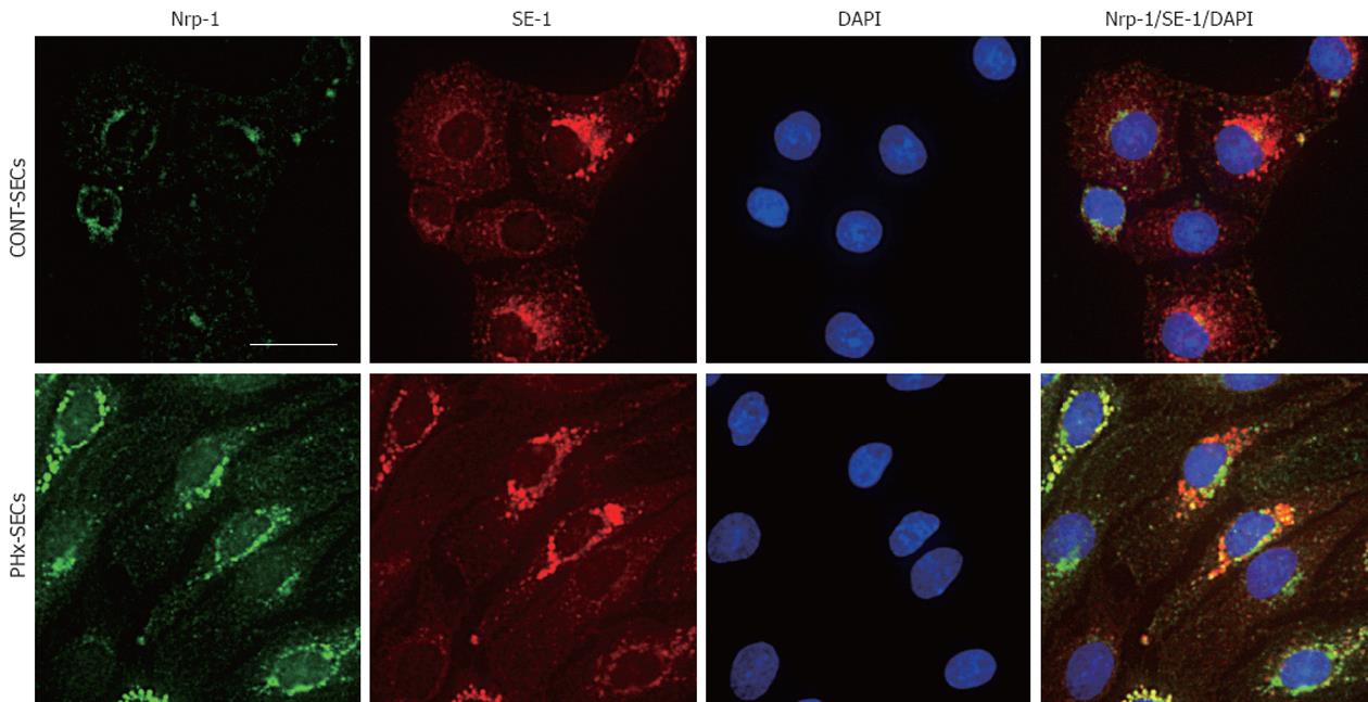
Figure 3 Immunofluorescent examination of neuropilin-1 and SE-1 in sinusoidal endothelial cells in primary culture.
A: Neuropilin-1 (Nrp-1) expression in sinusoidal endothelial cells (SECs) isolated from normal rats (CONT-SECs); B: Nrp-1 expression in SECs isolated from rats after partial hepatectomy (PHx-SECs). Green: Nrp-1; Red: SE-1; Blue: Nuclei stained with 4,6-diamidino-2-phenylindole dihydrochloride (DAPI). Scale bar, 20 μm.
Figure 4 Role of semaphorin 3A in sinusoidal endothelial cell migration and apoptosis.
A: Effect of semaphorin 3A (Sema3A) on migration of sinusoidal endothelial cells (SECs) isolated from normal rats (CONT-SECs) and rats after partial hepatectomy (PHx-SECs). SEC migration was assessed by cell transwell assay in the presence or absence of 5 nmol Sema3A for 24 h. Migration index represents the number of migratory cells/number of migratory cells in vehicle-treated PHx-SECs; B: Effect of Sema3A on apoptosis of CONT-SECs and PHx-SECs. Following incubation of SECs with 5 nmol Sema3A or vehicle for 24 h, apoptotic cells were determined by a terminal deoxynucleotidyl transferase-mediated deoxyuridine triphosphate nick end labeling method. Results are expressed as the mean ± SE. aP < 0.05, bP < 0.01 vs vehicle-treated PHx-SECs (n = 4).
- Citation: Fu L, Kitamura T, Iwabuchi K, Ichinose S, Yanagida M, Ogawa H, Watanabe S, Maruyama T, Suyama M, Takamori K. Interplay of neuropilin-1 and semaphorin 3A after partial hepatectomy in rats. World J Gastroenterol 2012; 18(36): 5034-5041
- URL: https://www.wjgnet.com/1007-9327/full/v18/i36/5034.htm
- DOI: https://dx.doi.org/10.3748/wjg.v18.i36.5034


