Copyright
©2012 Baishideng Publishing Group Co.
World J Gastroenterol. Sep 14, 2012; 18(34): 4798-4800
Published online Sep 14, 2012. doi: 10.3748/wjg.v18.i34.4798
Published online Sep 14, 2012. doi: 10.3748/wjg.v18.i34.4798
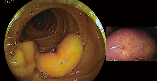
Figure 1 A 20 mm × 10 mm, white to yellowish, elongated, pedunculated jejunal polyp with erosions was detected in the patient following a double balloon enteroscopy.
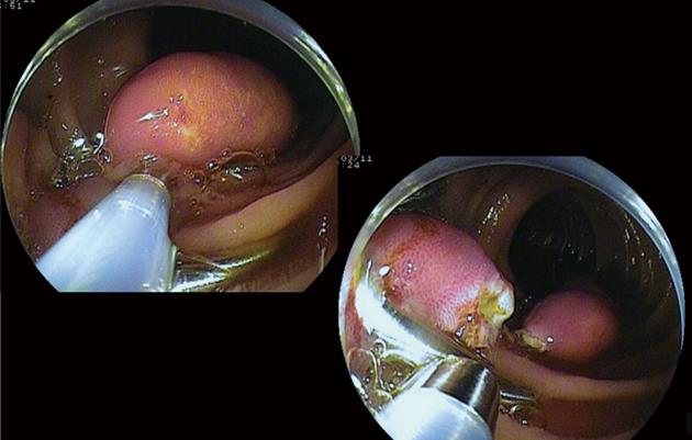
Figure 2 An endoscopic polypectomy was performed after the placement of two hemoclips at the polyp base.
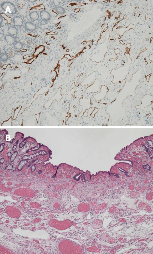
Figure 3 Histological view of the tumor revealing clusters of lymphatic vessels with a marked cystic dilatation in the submucosa and deep layer of the lamina propria mucosae.
A: Staining was performed with D2-40; B: Staining was performed with hematoxylin and eosin. These characteristics are consistent with the typical features of small-bowel lymphangioma with erosions.
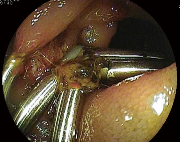
Figure 4 Lesion after treatment with hemoclipping and argon plasma coagulation.
- Citation: Kida A, Matsuda K, Hirai S, Shimatani A, Horita Y, Hiramatsu K, Matsuda M, Ogino H, Ishizawa S, Noda Y. A pedunculated polyp-shaped small-bowel lymphangioma causing gastrointestinal bleeding and treated by double-balloon enteroscopy. World J Gastroenterol 2012; 18(34): 4798-4800
- URL: https://www.wjgnet.com/1007-9327/full/v18/i34/4798.htm
- DOI: https://dx.doi.org/10.3748/wjg.v18.i34.4798


