Copyright
©2012 Baishideng Publishing Group Co.
World J Gastroenterol. Aug 28, 2012; 18(32): 4419-4426
Published online Aug 28, 2012. doi: 10.3748/wjg.v18.i32.4419
Published online Aug 28, 2012. doi: 10.3748/wjg.v18.i32.4419
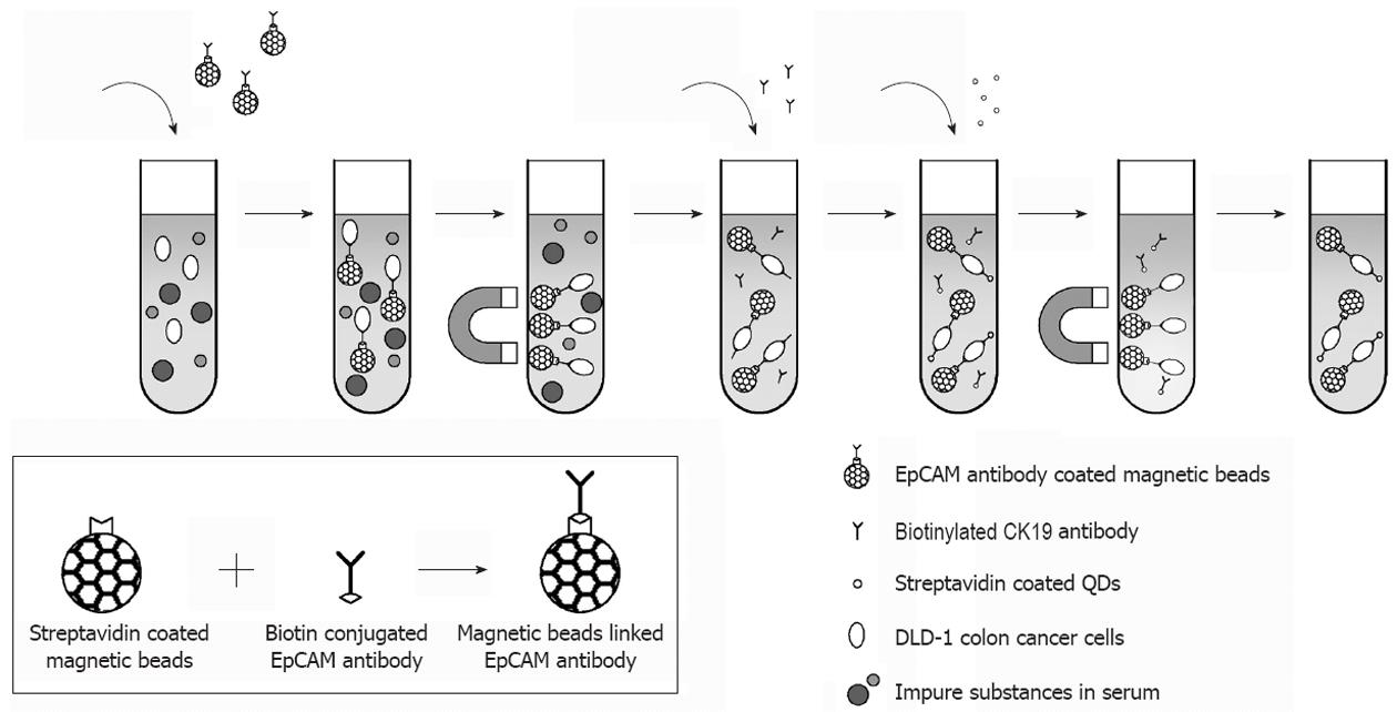
Figure 1 Circulating tumour cells are separated from the leucocytes using magnetic beads coupled with epithelial cell adhesion molecule antibody and a magnetic device.
These complexes are then tagged with cytokeratin-19 (CK19) biotinylated antibody and streptavidin-conjugated quantum dots (QDs) which lead to the detection of a fluorescent signal. EpCAM: Epithelial cell adhesion molecule; DLD-1: Human colon adenocarcinoma cell line.
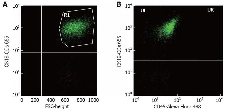
Figure 2 Representative fluorescence-activated cell sorting analysis of peripheral blood samples.
A: Events which fell within region R1 are confirmed to be cytokeratin 19 (CK19)+ and fluorescent in FL3 (anti-CK19 conjugated with QDs); B: From the R1 gate, the events confirmed to be CK19+CD45-are those in the UL region, in UR region the events represent those that are CK19+CD45+ and fluorescent in FL1 (anti-CD45 conjugated with Alexa Fluor). We deemed that the cells in the UL are those meeting the criteria for circulating tumor cells.
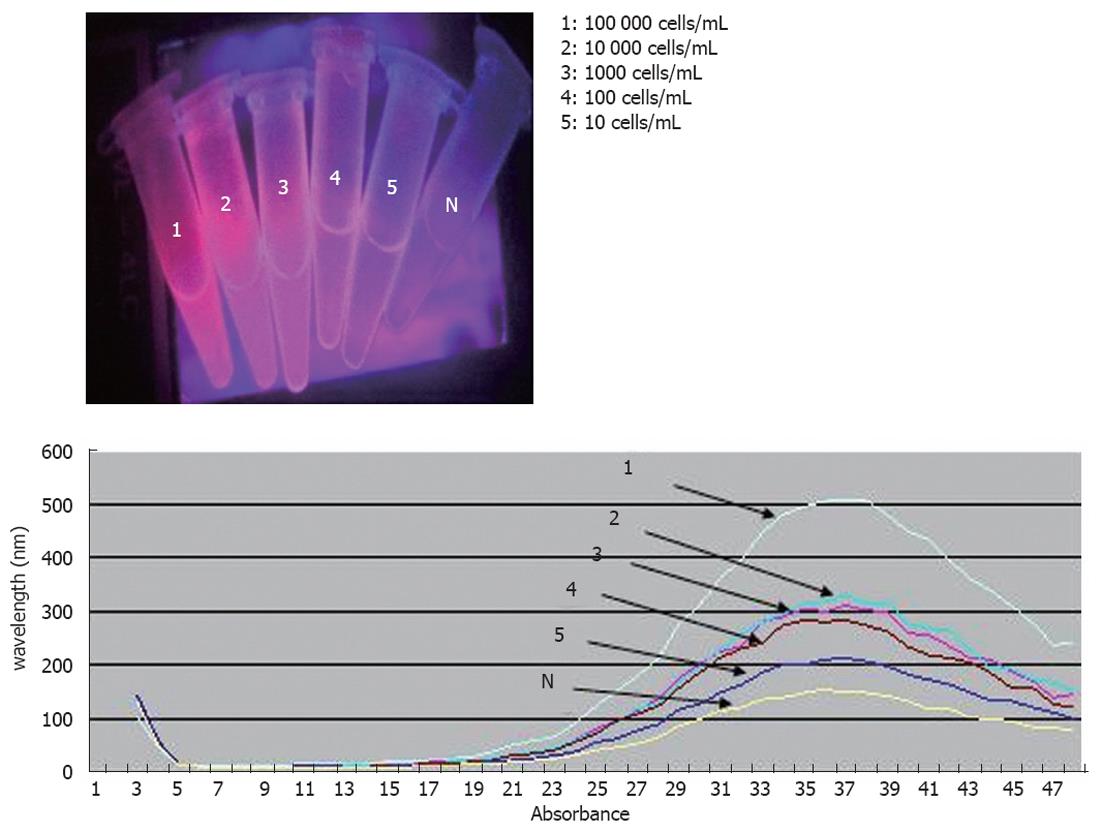
Figure 3 Representative results recorded by the proposed method for the human colon adenocarcinoma cell line serial dilutions ranging from 104 to 10 cells/mL (numbers 1-5) and negative controls.
Fluorescence is evident in the human colon adenocarcinoma cell line samples with concentration up to 10 cells/mL, as illustrated at the fluorescent emission spectra obtained after magnetic separation.
- Citation: Gazouli M, Lyberopoulou A, Pericleous P, Rizos S, Aravantinos G, Nikiteas N, Anagnou NP, Efstathopoulos EP. Development of a quantum-dot-labelled magnetic immunoassay method for circulating colorectal cancer cell detection. World J Gastroenterol 2012; 18(32): 4419-4426
- URL: https://www.wjgnet.com/1007-9327/full/v18/i32/4419.htm
- DOI: https://dx.doi.org/10.3748/wjg.v18.i32.4419


