Copyright
©2012 Baishideng Publishing Group Co.
World J Gastroenterol. Aug 21, 2012; 18(31): 4086-4090
Published online Aug 21, 2012. doi: 10.3748/wjg.v18.i31.4086
Published online Aug 21, 2012. doi: 10.3748/wjg.v18.i31.4086
Figure 1 Various sensors which are commercially available[12].
A: Miniature magnetometers offer orientation and acceleration information, [Bertda Services (SEA) Pte. Ltd.]; B: Three-dimensional (3-D) range camera used in a widely commercial product; C: 3-D guidance system used in endoscopy devices (inition).
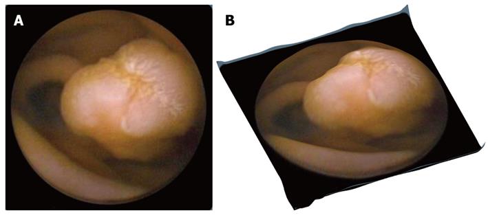
Figure 3 Original capsule endoscopy and 3-D represented image depicting a protrusion.
A: Original capsule endoscopy image depicting a protrusion (polyp); B: Its 3-D represented image processed by the proposed Shape from Shading approach.
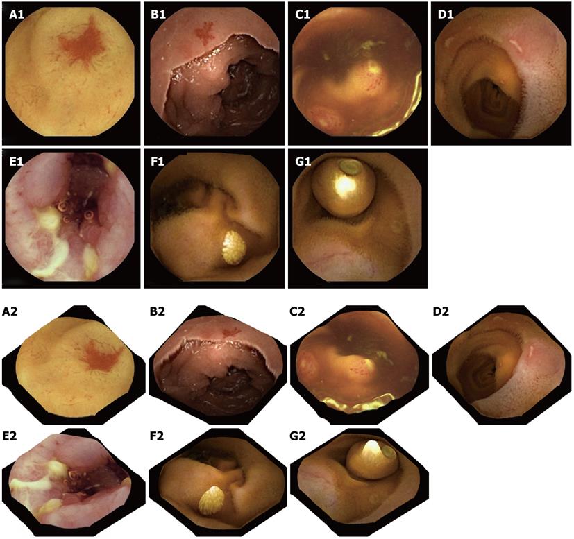
Figure 4 Two-dimensional capsule endoscopy images (upper panel) and three-dimensional representation of above structures is seen (lower panel).
A1, A2: P2 angioectasia; B1, B2: P1 angiectasia; C1, C2: Lymphoid hyperplasia with superficial ulceration; D1, D2: Aphthous ulcers; E1, E2: Serpiginous ulcers; F1, F2: Nodular lymphangiectasia; G1, G2: Another capsule endoscope still inside the small-bowel.
Figure 5 3-D representation of a single video capsule image with highlights removal (arrows).
- Citation: Koulaouzidis A, Karargyris A. Three-dimensional image reconstruction in capsule endoscopy. World J Gastroenterol 2012; 18(31): 4086-4090
- URL: https://www.wjgnet.com/1007-9327/full/v18/i31/4086.htm
- DOI: https://dx.doi.org/10.3748/wjg.v18.i31.4086


