Copyright
©2012 Baishideng Publishing Group Co.
World J Gastroenterol. Aug 14, 2012; 18(30): 3992-3996
Published online Aug 14, 2012. doi: 10.3748/wjg.v18.i30.3992
Published online Aug 14, 2012. doi: 10.3748/wjg.v18.i30.3992
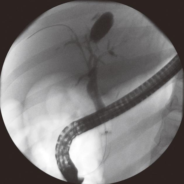
Figure 1 Ex vivo and in vivo views of the anchoring balloon.
A: Overall view of the anchoring balloon; B: Fluoroscopic view of an inflated anchoring balloon that has been passed over a guidewire and anchored in one of the intrahepatic bile ducts; C: Fluoroscopic view of an ultraslim upper endoscope that has been backloaded over the catheter of an inflated anchoring balloon and advanced into the bile duct to the level of the confluence of the right and left hepatic ducts.
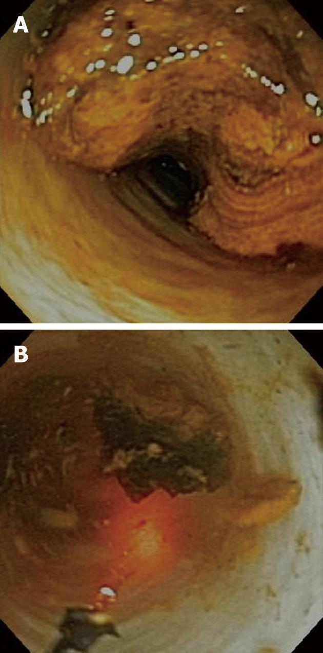
Figure 2 Cholangioscopic view of a large stone.
A: The stone could not be removed during prior endoscopic retrograde cholangiopancreatography. A plastic stent was placed and the patient was referred for cholangioscopy-guided laser lithotripsy. Impression of the plastic stent in the stone is clearly visible; B: Cholangioscopic view of the same stone after laser lithotripsy. The laser probe is seen in the left lower corner of the picture.
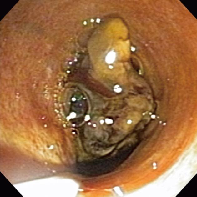
Figure 3 Cholangioscopic image of a tumor in the proximal common hepatic duct.
The anchoring balloon catheter is seen in the left lower corner of the picture.
Figure 4 Biloma at the site of the anchored balloon within an intrahepatic bile duct.
- Citation: Parsi MA, Stevens T, Vargo JJ. Diagnostic and therapeutic direct peroral cholangioscopy using an intraductal anchoring balloon. World J Gastroenterol 2012; 18(30): 3992-3996
- URL: https://www.wjgnet.com/1007-9327/full/v18/i30/3992.htm
- DOI: https://dx.doi.org/10.3748/wjg.v18.i30.3992


