Copyright
©2012 Baishideng Publishing Group Co.
World J Gastroenterol. Jul 28, 2012; 18(28): 3770-3774
Published online Jul 28, 2012. doi: 10.3748/wjg.v18.i28.3770
Published online Jul 28, 2012. doi: 10.3748/wjg.v18.i28.3770
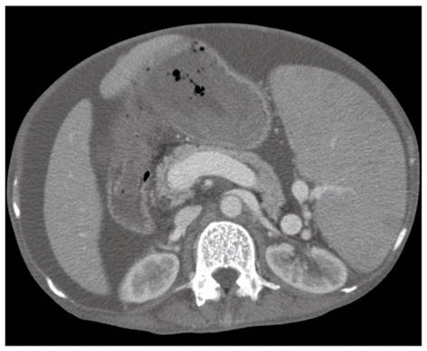
Figure 1 Spleen enlargement and dilatation of the splenic vein on abdominal computed tomography.
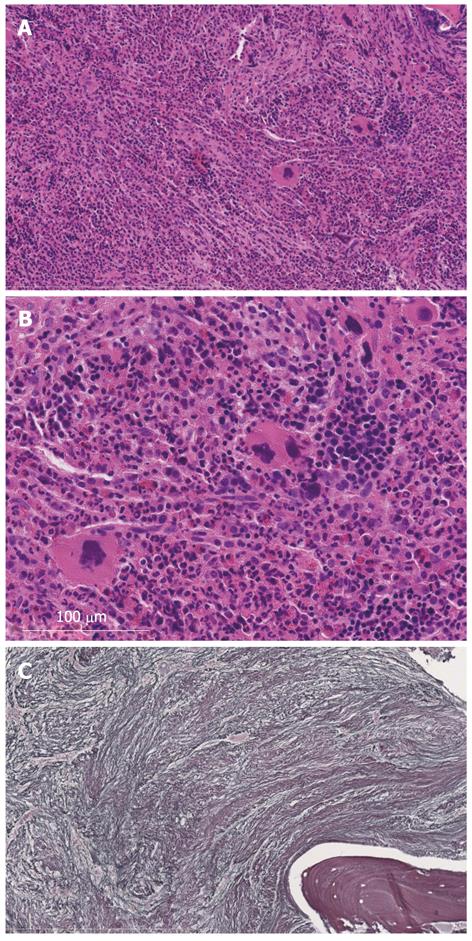
Figure 2 Microscopic findings on autopsy on bone marrow biopsy.
A: Hematoxylin and eosion, × 20; B: Hematoxylin and eosion, × 40; C: Silver, × 40. Adipocyte disappearance and markedly decreased hematopoietic cells.
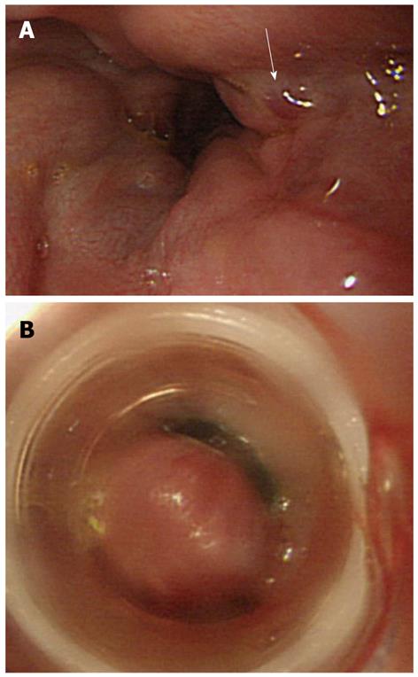
Figure 3 Endoscopic image in the lower esophagus.
A: Showing a hematocystic spot by the white arrow; B: Endoscopic variceal ligation was thus conducted.
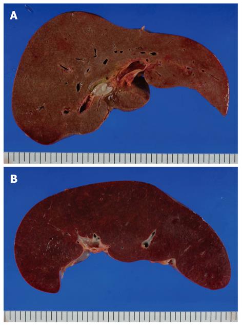
Figure 4 Macroscopic findings on autopsy.
A: The liver weighed 1856 g; B: The spleen 1572 g indicating hepatosplenomegaly.
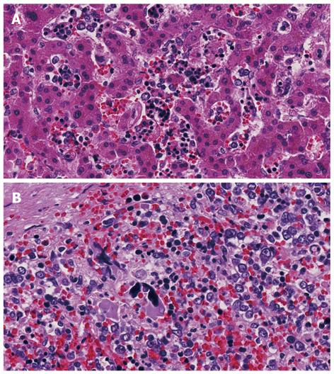
Figure 5 Microscopic findings on autopsy stained with hematoxylin and eosion.
A: Liver; B: Spleen. Both the liver and the spleen showed extramedullary hemopoiesis.
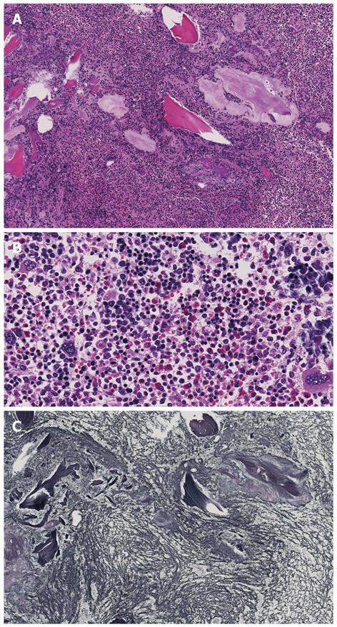
Figure 6 Microscopic findings on autopsy (bone marrow).
A: Hematoxylin and eosion (HE), × 10; B: HE, × 40; C: Silver, × 10. Adipocyte disappearance and 3-lineage differentiation were seen. Silver staining showed severe fibrosis with mainly argyrophilic fibers.
- Citation: Tokai K, Miyatani H, Yoshida Y, Yamada S. Multiple esophageal variceal ruptures with massive ascites due to myelofibrosis-induced portal hypertension. World J Gastroenterol 2012; 18(28): 3770-3774
- URL: https://www.wjgnet.com/1007-9327/full/v18/i28/3770.htm
- DOI: https://dx.doi.org/10.3748/wjg.v18.i28.3770


