Copyright
©2012 Baishideng Publishing Group Co.
World J Gastroenterol. Jul 28, 2012; 18(28): 3696-3704
Published online Jul 28, 2012. doi: 10.3748/wjg.v18.i28.3696
Published online Jul 28, 2012. doi: 10.3748/wjg.v18.i28.3696
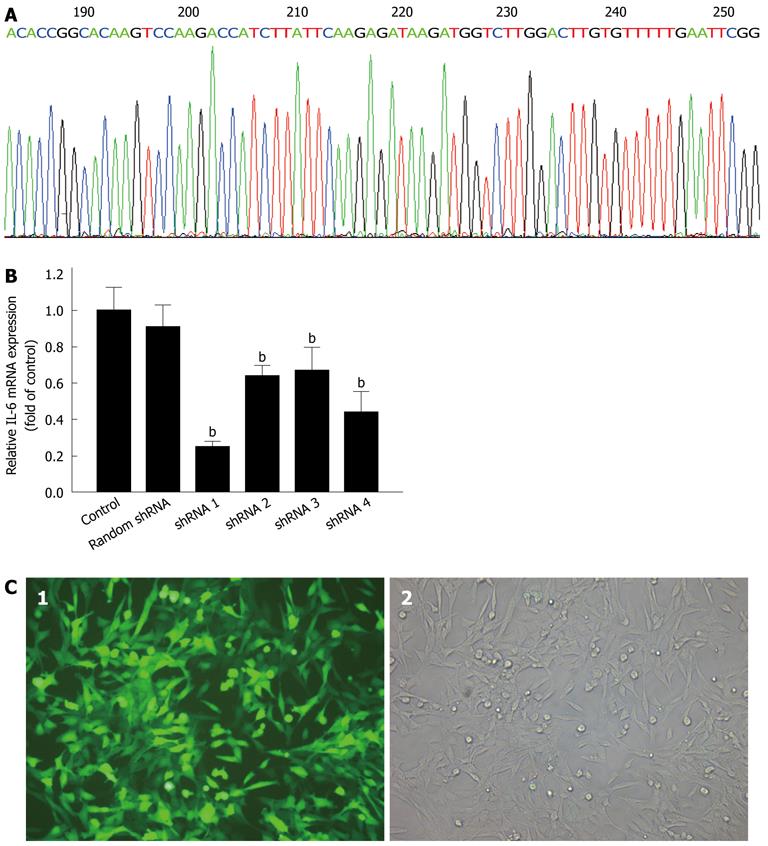
Figure 1 Short hairpin RNA design, interference efficiency assay, and lentivirus transduction.
A: DNA sequence analysis of pGCSIL/Interleukin-17A receptor (IL-17RA) short hairpin RNA (shRNA) 1; B: Lentiviral-mediated IL-17RA shRNA 1, 2, 3, and 4 (shRNA 1, 2, 3, and 4) inhibited IL-17RA mRNA expression in hepatic stellate cells (HSCs). HSCs infected with lentivirus at a multiplicity of infection of 10 for 72 h, and IL-17RA mRNA expression was quantified by polymerase chain reaction. IL-17RA shRNA 1 was the most efficient silencing tool for IL-17RA. bP < 0.01 vs control; C: Cell morphology of HSCs under fluorescence (1) and light (2) microscope after 72 h transduction.
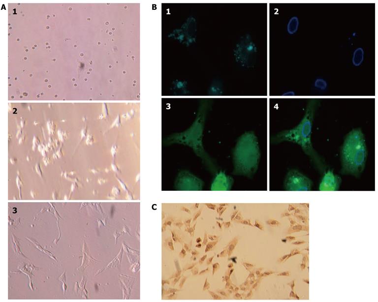
Figure 2 Isolation, culture, and identification of primary rat hepatic stellate cells.
A: Primary rat hepatic stellate cells (HSCs) on days 1, 2, and 5 after isolation and plated in culture; B: HSCs isolated from Sprague-Dawley rats showed characteristic multiple lipid droplets with a rapid fade of the blue-green autofluorescence at 328 nm. 1: Lipid droplets; 2: Cell nucleus; 3: Cell body. 4: Merged images; C: HSC culture after the first passage and stained with anti-α-smooth muscle actin antibody.
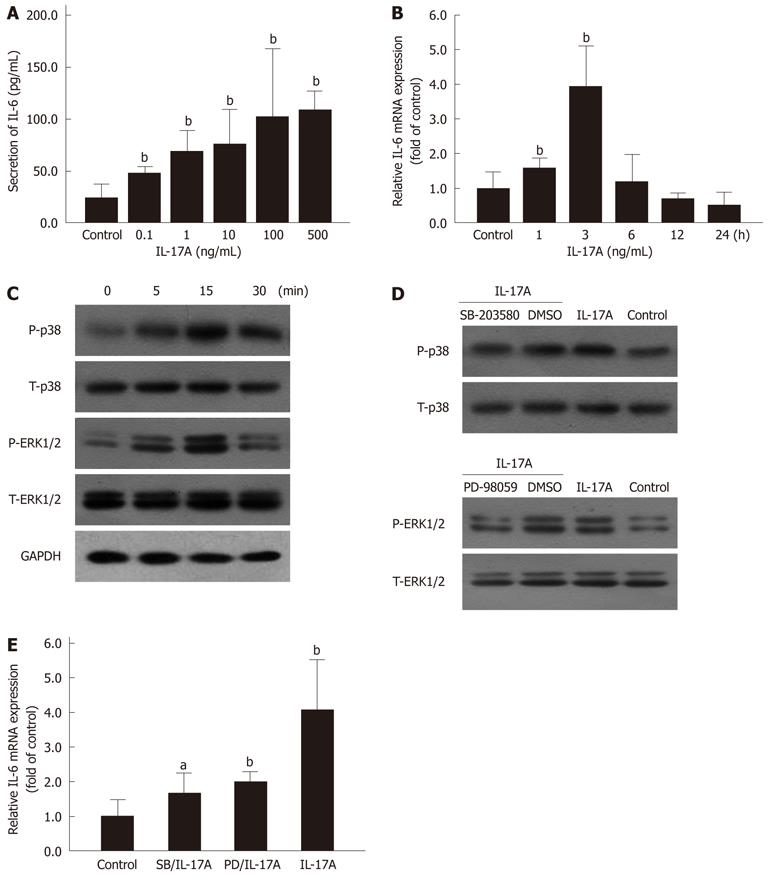
Figure 3 Mitogen activated protein kinases pathway involved in interleukin 17A induced interleukin 6 expression.
A: Secretion of interleukin (IL)-6 in hepatic stellate cells (HSCs) induced by interleukin 17A (IL-17A) was determined using enzyme-linked immunosorbent assay. bP < 0.01 vs control; B: IL-6 mRNA expression in HSCs induced by IL-17A (100 ng/mL) was measured by polymerase chain reaction. bP < 0.01 vs control; C: Phosphorylation of p38 MAPK and ERK1/2 induced by IL-17A (100 ng/mL) in HSCs was detected using Western blotting; D: Phosphorylation of p38 MAPK and ERK1/2 induced by IL-17A (100 ng/mL) for 15 min was blocked by preincubation with MAPKs inhibitors, SB-203580 (1 μmol/L in DMSO, 30 min) and PD-98059 (10 μmol/L in DMSO, 1 h); E: IL-6 mRNA expression induced by IL-17A (100 ng/mL) for 3 h exposure was inhibited by preincubation with SB-203580 (SB) and PD-98059 (PD) for the same time period as above. aP < 0.05, bP < 0.01 vs control. T: Total; P: Phosphorylation; GAPDH: Glyceraldehyde-3-phosphate dehydrogenase; MAPK: Mitogen activated protein kinases; ERK: Extracellular regulated protein kinases.
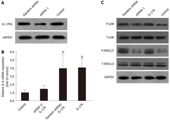
Figure 4 Lentiviral-mediated interleukin 17A receptor short hairpin RNA arrested interleukin 6 expression, partly through suppressing phosphorylation of p38 mitogen activated protein kinases and extracellular regulated protein kinases 1/2.
A: Lentiviral-mediated interleukin (IL)-17A receptor short hairpin RNA (shRNA) 1 [at multiplicity of infection (MOI) = 10 for 72 h] inhibited the expression of IL-17RA in hepatic stellate cells (HSCs); B: Lentiviral-mediated IL-17RA shRNA 1 inhibited IL-17A-induced IL-6 mRNA expression in HSCs. HSCs were infected with IL-17RA shRNA 1 or random shRNA lentivirus at MOI = 10 for 72 h, and followed by 3 h exposure to IL-17A (100 ng/mL). bP < 0.01 vs control; C: Lentiviral-mediated IL-17RA shRNA 1 inhibited phosphorylation of p38 MAPK and ERK1/2. HSCs were transduced with IL-17RA shRNA 1 or random shRNA lentiviral at MOI = 10 for 72 h, and followed by 15 min exposure to IL-17A (100 ng/mL). T: Total, P: Phosphorylation; GAPDH: Glyceraldehyde-3-phosphate dehydrogenase.
- Citation: Zhang SC, Zheng YH, Yu PP, Min TH, Yu FX, Ye C, Xie YK, Zhang QY. Lentiviral vector-mediated down-regulation of IL-17A receptor in hepatic stellate cells results in decreased secretion of IL-6. World J Gastroenterol 2012; 18(28): 3696-3704
- URL: https://www.wjgnet.com/1007-9327/full/v18/i28/3696.htm
- DOI: https://dx.doi.org/10.3748/wjg.v18.i28.3696


